Drei Posterbeiträge zu TIGIT vom Annual Meeting der American Association for Cancer Research (AACR), Chicago, sind hier mit ihren Originalankündigungen dokumentiert und als Original-PDFs abrufbar. Die Ankündigungstexte liegen im englischen Original vor.
Ankündigung · 15.05.2018
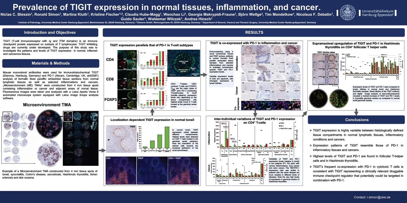
Prevalence of TIGIT expression in normal tissues, inflammation, and cancer
American Association of Cancer Research (AACR) Annual Meeting, Chicago, Apr 14-18, 2018 – Poster presentation:
Mouse monoclonal antibody TG1 was used for immunohistochemical analysis of routine formalin fixed paraffin embedded tissue sections from normal lymphatic tissues as well as selected inflamations and cancers and compared to expression patterns of PD1.
Data provided by: Institue of Pathology, University Medical Center Hamburg-Eppendorf, Martinistr. 52, 20246 Hamburg, Germany. Dianova GmbH, Warburgstr. 54, 20354 Hamburg, Germany. Department of General, Visceral and Thoracic Surgery, University Medical Center Hamburg-Eppendorf, Martinistr. 52, 20246 Hamburg, Germany.
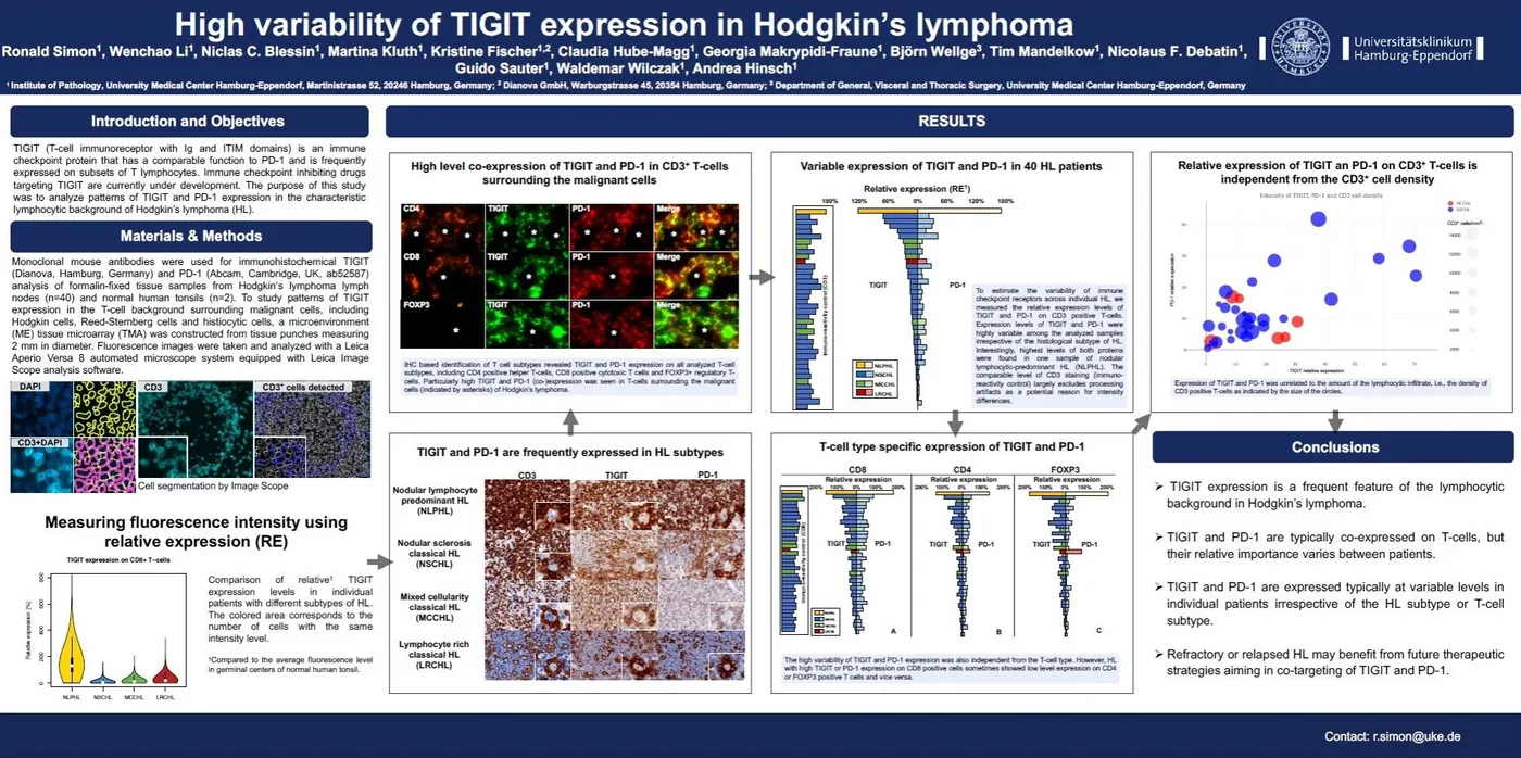
High variability of TIGIT expression in Hodgkin’s lymphoma
American Association of Cancer Research (AACR) Annual Meeting, Chicago, Apr 14-18, 2018 – Poster presentation:
To study patterns of TIGIT expression in the T-cell background surrounding malignant cells, including Hodgkin cells, Reed-Sternberg cells and histiocytic cells, a microenvironment tissue microarray (TMA) was constructed. Immunofluorescence studies with monoclonal antibody TG1 have been perfomed to analyse T-cell type specific expression of TIGIT.
Data provided by: Institue of Pathology, University Medical Center Hamburg-Eppendorf, Martinistr. 52, 20246 Hamburg, Germany. Dianova GmbH, Warburgstr. 54, 20354 Hamburg, Germany. Department of General, Visceral and Thoracic Surgery, University Medical Center Hamburg-Eppendorf, Martinistr. 52, 20246 Hamburg, Germany.
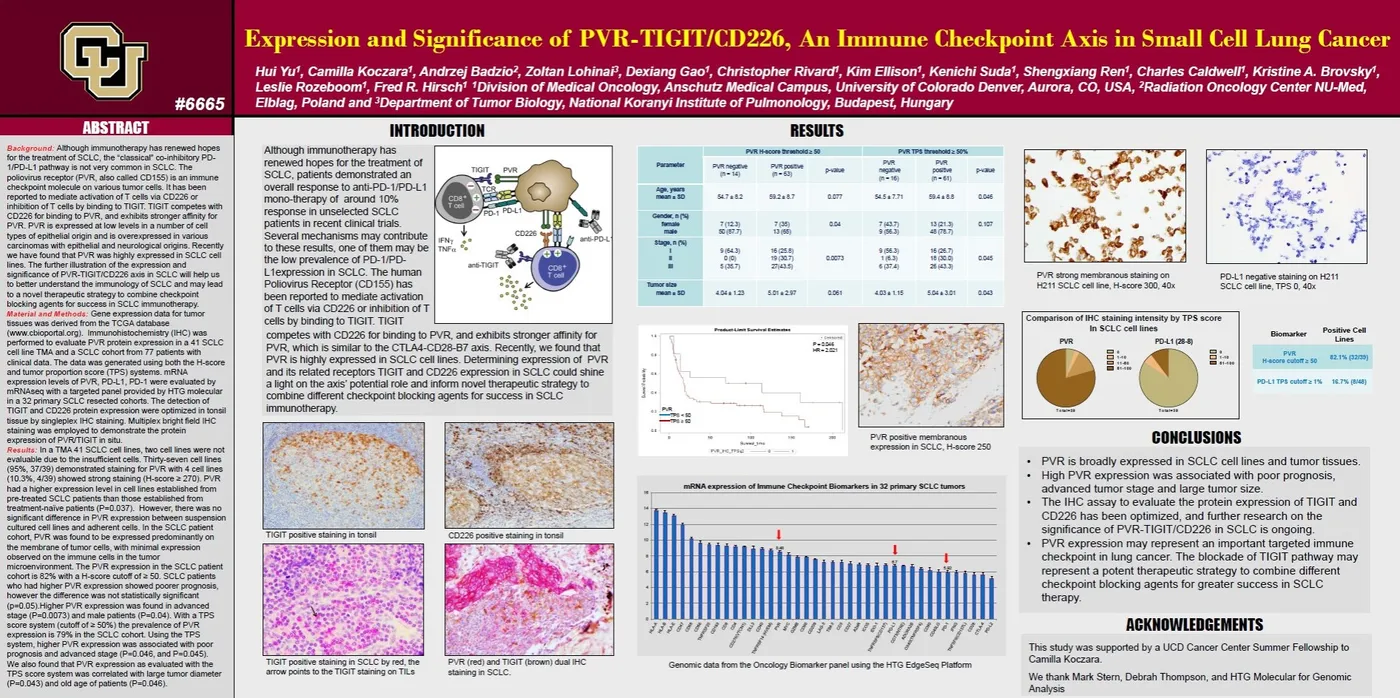
Expression and Significance of PVR-TIGIT/CD226 in Small Cell Lung Cancer
American Association of Cancer Research (AACR) Annual Meeting, Chicago, Apr 14-18, 2018 – Poster presentation:
The “classical” co-inhibitory PD-1/PD-L1 pathway is not very common in SCLC, but IHC-analysis of the expression and significance of PVR (CD155)-TIGIT/CD226 axis in SCLC highlights a potential role for checkpoint blockade of the TIGIT pathway and may represent a potent therapeutic strategy in SCLC therapy.
Data provided by: Hirsch Biomarker Analysis Laboratory, Divison of Medical Oncology, Anschutz Medical Campus, University of Colorado Denver, Aurora, CO, USA. Radiation Oncology Center NU-Med, Elblag, Poland. Department of Tumor Biology, National Kuranyi Institute of Pulmonology, Budapest, Hungary.
Kongressposter sind Beiträge der auf dem jeweiligen Poster genannten Arbeitsgruppen; Aussagen zu therapeutischen Strategien geben die Forschungsperspektive der Posterautoren zum TIGIT-Signalweg wieder. ONCOdianova-Antikörper: Nur für Forschungszwecke. Nicht zur Verwendung in diagnostischen Verfahren.