TG1 — a published anti-TIGIT antibody for human FFPE IHC.

Clone TG1 · Cat. no. DIA-TG1-M · RRID AB_3750391

TIGIT is expressed on T cells in tumors. The ultra-validated clone TG1 allows the identification of TIGIT-positive T cells in human tumor tissue sections (FFPE). The new clone TG2 shows a stronger staining intensity.

Scientific USP

12 clone-specific peer-reviewed publications

1,778Evaluable tumors
86Tumor entities
  • Brightfield + fluorescence multiplex IHC
  • WB · ELISA · TIGIT-protein pre-absorption

Clone TG1 has been validated on large quantities of tumor tissues and beta-tested by different labs for IHC-FFPE. With antibody clone TG1, TIGIT could for the first time be detected immunohistochemically in tumor tissue.

Full list with DOI and PMID: publications.

Mouse monoclonal antibody for studying TIGIT in human FFPE tissue. Clone TG1 is documented by twelve peer-reviewed references, covering brightfield IHC, automated platforms and fluorescence multiplex IHC.

  • CloneTG1 (monoclonal)
  • Catalog numberDIA-TG1-M
  • RRIDAB_3750391
  • Species reactivityHuman
  • Host speciesMouse
  • IsotypeMouse IgG1/κ
  • ApplicationsIHC on FFPE tissue · Western blot
  • IHC starting dilution1:50 – 1:150
  • Positive controlTonsil
  • Staining patternMembranous (lymphocyte subsets)
  • Format100 µl, lyophilized

Complete product data: Description, section P·05 ↓

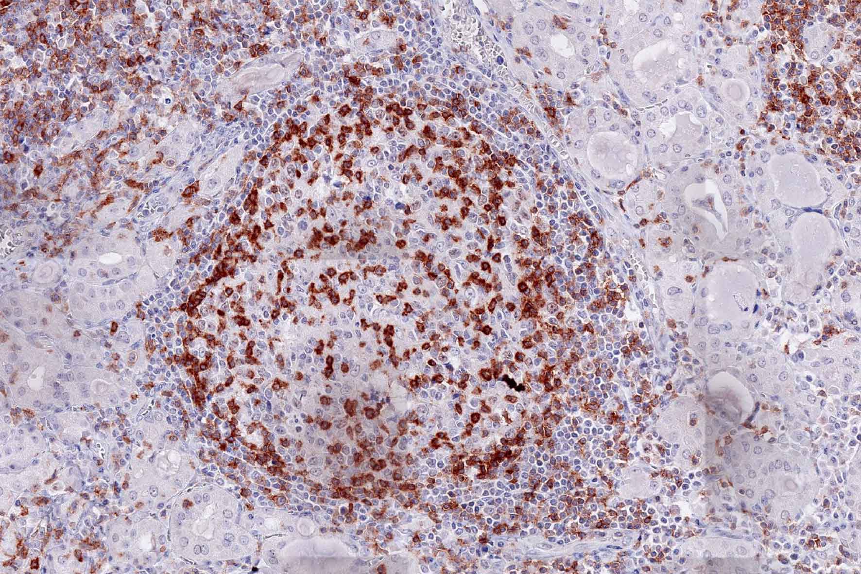
Product image of the ONCOdianova anti-TIGIT antibody clone TG1: TIGIT immunohistochemistry of an inflammatory lymphoid infiltrate in Hashimoto thyroiditis
Product image · Clone TG1 · DIA-TG1-M
Evidence layer 01

Clone-specific publications

TG1 is documented by twelve peer-reviewed references — including Hodgkin lymphoma, seminoma, lung cancer, gallbladder cancer, colorectal cancer, melanoma, uveal melanoma, cutaneous squamous cell carcinoma and Merkel cell carcinoma. Three AACR congress presentations on TIGIT are documented in section P·12.

Evidence layer 02

Broad FFPE documentation

A systematic study evaluated 1,778 tumors representing 86 tumor entities and also documented TG1 in normal and inflammatory tissues, with Western blotting, ELISA and pre-absorption reported as specificity assessments.

Evidence layer 03

Manual, automated and multiplex

Documented manual and Leica Bond RX starting protocols; independent studies report Dako Autostainer Link 48, Agilent Omnis and fluorescence multiplex IHC. Conditions must be validated in the user's laboratory.

IHC image gallery and ONCOdianova reference images: section P·03 ↓

Antibody clones TG1 and TG2 for detecting TIGIT-positive T cells in FFPE tissue.

TIGIT at the Immune Checkpoint

A growing number of immune checkpoints are emerging as targets for anticancer therapy. Cancer immunology studies have shown that cancer cells, together with cells of the surrounding microenvironment, generate co-inhibitory signals by upregulating the expression of components that suppress the antitumor immune response. The TIGIT pathway interacts with different inhibitory checkpoint pathways. TIGIT provides significant promise for immunotherapy, especially in combination with other immune checkpoint inhibitors.

The immunoreceptor TIGIT (T-cell immunoreceptor with Ig and ITIM domains) is a member of the poliovirus receptor (PVR) family. Expression of TIGIT has been reported on NK cells, regulatory T cells, follicular T helper cells, memory CD4+ T cells, and CD8+ T cells, but TIGIT is not expressed on B cells or naive CD4+ T cells. TIGIT acts as an inhibitory immune checkpoint on both T cells and natural killer (NK) cells by a highly complex pathway. Known ligands for TIGIT include CD155 and CD112. Moreover, the TIGIT/CD155/CD112 network also interacts with further checkpoint regulators.

In inflammation and in multiple cancer models, T cells have been shown to upregulate TIGIT expression. In several types of cancer, the ligands CD155 and CD112 are also highly expressed on dendritic cells and macrophages. Moreover, TIGIT expression is highly correlated with the expression of other co-inhibitory molecules, including PD-1. In addition to directly inhibiting cytotoxic T-cell activity, TIGIT can stimulate an immunosuppressive microenvironment by influencing other immune cells: for example, TIGIT binds CD155 on the surface of dendritic cells or manipulates NK cell activity. Drugs inhibiting TIGIT activity are currently being developed.

New – clone TG2

The newly developed clone TG2 has a higher affinity to the target protein TIGIT and shows a stronger staining intensity in IHC studies on tonsil when compared to TG1.

  • Higher affinity as compared to clone TG1 (tested on tonsil)
  • Stronger staining intensity
  • Higher dilutability

TG1 and TG2 are separate clones with their own product data and catalog numbers. Product data and publications for TG2 are on the TG2 page. NCBI-verified TIGIT cell biology with clone TG1 in human FFPE tissue is documented below in the TIGIT in research section.

Everything about DIA-TG1-M, complete in eight sections.

Data package.

Datasheet

DIA-TG1-M

Product data, instructions for use, references and figures A–D.

Download PDF ↓

PDF · V17 · 25 Feb 2025 · 272 KB

Tech note

TIGIT staining

Manual and Leica Bond RX staining procedures, tonsil (100×/200×/400×), retrieval comparison and Western blot figures.

Download PDF ↓

PDF · V1 · Nov 2019 · 988 KB

Controlled product data.

ProductAnti-TIGIT (Human) from Mouse
Catalog numberDIA-TG1-M (earlier presentation of the same clone: DIA-TG1, 200 µg)
Distributor catalog numberODN-DIA-TG1-M (BIOZOL)
SKUTIGIT
Product categoryCancer Immunology IHC marker
SpecificityTIGIT
TargetTIGIT — T-cell immunoreceptor with Ig and ITIM domains (Gene ID 201633, UniProt Q495A1)
Species reactivityHuman
Host speciesMouse
IsotypeMouse IgG1/κ
CloneTG1 (monoclonal)
RRIDAB_3750391
ImmunogenRecombinant peptide from the extracellular domain of human TIGIT
ApplicationsIHC on FFPE tissue; Western blot
ConjugationUnconjugated
IHC starting range1:50 – 1:150 — 1:50 for biotin/(strept)avidin-based detection, 1:100–1:150 for polymer-based detection; optimal dilution to be determined by the user for tissue, fixation, platform and detection system
FormatLyophilized; antibody purified from culture supernatant
Quantity100 µl
ReconstitutionRestore to 100 µl with sterile distilled water; gentle shaking for 10 minutes
Buffer / formulationPBS, pH 7.4, 2% BSA, 0.05% sodium azide
BSA-free variantBSA-free high-concentration variant available on request
Epitope retrievalHeat-induced epitope retrieval required; autoclave 121 °C, 5 min, Tris-EDTA-citrate pH 7.8 recommended
Positive controlTonsil
Staining patternMembranous (lymphocyte subsets)
StorageLyophilized at 2–8 °C; long-term at −20 °C (stable for at least one year); reconstituted at 2–8 °C short term (several weeks); avoid repeated freeze/thaw cycles
Associated antibodiesDIA-TG2-M (anti-TIGIT, clone TG2) · DIA-R12 (anti-PVRIG/CD112R, clone R12) — as listed in the datasheet
Notice of useFor Research Use Only. Not for use in diagnostic procedures.
ManufacturerONCOdianova GmbH

The technical data above are taken from datasheet V17 (25 Feb 2025) and tech note V1 (Nov 2019). RRID, gene and protein entries link to their registries; published staining conditions are documented in the publications section. The data apply to the current DIA-TG1-M product (100 µl). Published dilutions range from 1:25 to 1:150 depending on study and method; each laboratory must validate its own conditions.

Reactivity of clone TG1.

Reactivity

Clone TG1 is the first monoclonal antibody detecting TIGIT in routine formalin-fixed paraffin-embedded (FFPE) tissues. TG1 has been validated for the detection of TIGIT-positive T cells with a focus on tumor infiltrating T cells (TILs). Anti-TIGIT clone TG1 allows the identification of TIGIT in the tumor microenvironment under pathological conditions.

TIGIT (T-cell immunoreceptor with Ig and ITIM domains) is a member of the poliovirus receptor (PVR) family. TIGIT is expressed on subsets of T lymphocytes and controls an immune checkpoint. The expression of TIGIT has been reported on NK cells, regulatory T cells, follicular T helper cells, memory CD4+ T cells, and CD8+ T cells, but TIGIT is not expressed on B cells or naive CD4+ T cells. Naive CD4+ T cells may upregulate TIGIT expression upon activation. In many different cancer models, TIGIT has been shown to be upregulated on T cells. The ligands CD155 and CD112 are also highly expressed on dendritic cells and macrophages in different cancer diseases. Moreover, TIGIT expression is highly correlated with the expression of further co-inhibitory molecules like PD-1. In addition to its direct inhibitory effect on cytotoxic T cells, TIGIT can stimulate an immunosuppressive microenvironment by influencing other immune cells: for example, TIGIT can bind CD155 on the surface of dendritic cells or is able to manipulate NK cell activity. TIGIT inhibiting drugs are currently being developed. Immunohistochemical (IHC) application of monoclonal antibody TG1 may provide valuable information for clinical research and potential therapeutic interventions specifically targeting TIGIT at its cancer immunology checkpoint.

Antibody clone TG1

Clone TG1 is the first monoclonal antibody for the immunohistochemical (IHC) detection of TIGIT (T-cell immunoreceptor with Ig and ITIM domains) in routine FFPE human tissue specimens. Furthermore, TG1 has been validated for the identification of TIGIT-positive T cells infiltrating human tumors, with the aim of allowing detection of TIGIT in the tumor microenvironment by IHC. Immunohistochemical (IHC) application of monoclonal antibody TG1 may provide valuable information for clinical research and potential therapeutic interventions specifically targeting the TIGIT-related tumor immunology checkpoint.

Species reactivityHuman
Host speciesMouse
Detected with TG1 in FFPE tissueCD8+ cytotoxic T cells, CD4+ T-helper cells, FOXP3+ regulatory T cells and NK cells
Positive controlTonsil
Staining patternMembranous, lymphocyte subsets
ApplicationsIHC on human FFPE tissue; Western blot

IHC protocols and published applications.

ONCOdianova application protocol

Manual FFPE IHC — ONCOdianova protocol

Clone
TG1 · DIA-TG1 / DIA-TG1-M
Antigen retrieval
Autoclave · 121 °C · 5 min · TEC buffer pH 7.8
Dilution
1:50–1:150
Primary incubation
1 h · 37 °C · moist chamber
Detection
DAKO EnVision Polymer-HRP mouse/rabbit
Chromogen
DAB

Source: ONCOdianova Technical Note V01 “Anti-TIGIT / DIA-TG1 · clone TG1” — full document under Documents. The automated Leica Bond RX procedure of the same Technical Note is kept separate in procedure 2.

Peer-reviewed · with ONCOdianova involvement

Published brightfield IHC application — seminoma

Antibody
DIA-TG1 · mouse monoclonal
Dilution
1:70
Antigen retrieval
Autoclave · 121 °C · 5 min · pH 7.8 buffer
Primary incubation
60 min · 37 °C
Detection
EnVision Kit (Dako / Agilent)
Material
78 seminomas · FFPE tissue microarray

Hinsch A. et al., Oncol Lett 2019;18(2):1497–1502 · doi:10.3892/ol.2019.10428

Peer-reviewed · with Dianova involvement

Published multiplex fluorescence IHC

Antibody
Dianova #DIA-TG1
Dilution
1:150
Antigen retrieval
pH 9
Staining order
2nd
Dye
Opal 570
Scope of the study
> 1,700 tumour samples from 86 tumour entities; TIGIT and/or PD-1 by brightfield and/or multiplex fluorescence IHC

Blessin N.C. et al., Dis Markers 2019:5160565 · doi:10.1155/2019/5160565 — the same study reports pH 7.8 and 1:70 for the brightfield procedure; the multiplex values do not apply to brightfield.

Independent third-party application

Published application — Leica BOND-III

Antibody
DIA-TG1
Dilution
1:25
Platform
BOND-III Automated Immunostainer (Leica Microsystems)
Antigen retrieval
BOND Epitope Retrieval Solution 2 · 30 min
Primary incubation
30 min
Detection
BOND Polymer Refine Detection Kit · DAB
Material
4-µm FFPE sections · 100 consecutive colorectal cancer cases

Murakami D. et al., PLOS ONE 2022;17(3):e0265908 · doi:10.1371/journal.pone.0265908 — application reported by the research group, not an ONCOdianova protocol recommendation.

Independent third-party application

Published application — melanoma research

Antibody
DIA-TG1 · mouse monoclonal
Dilution
1:50
Platform
Dako Omnis (Dako / Agilent)
Antigen retrieval
Target retrieval solution pH 6 · 10 min · 117 °C · steam pressure cooker
Primary incubation
overnight at 4 °C
Detection
Alkaline Phosphatase Red Detection Kit
Positive control
Tonsillar tissue
Material
4-µm paraffin sections

Niebel D. et al., Clin Epigenetics 2022;14:50 · doi:10.1186/s13148-022-01270-2 — application reported by the research group, not an ONCOdianova protocol recommendation.

The protocols and published workflows shown here represent documented application examples. Antibody dilution, antigen retrieval, detection chemistry and staining conditions may require laboratory-specific optimization and validation.

Documented starting protocols. Both procedures come from Tech Note V1 (November 2019) for clone TG1. The stock concentration of 0.4 mg/ml and the reconstitution in 500 µl stated there belong to the DIA-TG1 format (200 µg); DIA-TG1-M (100 µl) is reconstituted in 100 µl. Dilutions of 1:50 – 1:150 are starting values; each laboratory validates tissue, fixation, platform and detection itself.

Procedure 1Manual immunostaining · IHC on FFPE tissueTech Note V1 · 11/2019

Slide preparation

  1. Mount 4 µm sections on Superfrost slides
  2. Deparaffinize tissue sections in xylene (2 × 5 min)
  3. Rehydrate in descending ethanol series (100%, 90%, 80%, 70%)
  4. Rinse 5 min in TBST* buffer

Pretreatment (epitope retrieval)

  1. Autoclave for 5 min** at 121 °C in TEC*** buffer pH 7.8 (1× concentration)
  2. Rinse 5 min in TBST* buffer

Peroxidase blocking

  1. Incubate 10 min in Peroxidase-Blocking Solution (#S2023, DAKO REAL™)
  2. Rinse 2 × 5 min in TBST* buffer

Antibody incubation

  1. Dilute primary antibody (TG-1, stock 0.4 mg/ml, Tech Note V1) 1:50 – 1:150 in antibody diluent (#S2022, DAKO REAL™)
  2. Cover tissue section with 100–200 µl diluted antibody
  3. Incubate 1 h at 37 °C in a moist chamber
  4. Rinse 2 × 5 min in TBST* buffer
  5. Apply DAKO EnVision Polymer-HRP mouse/rabbit kit (#K5007, DAKO REAL™) according to the kit protocol
  6. Rinse 2 × 5 min in TBST* buffer

Chromogen and counterstain

  1. Cover slides for 10 min with DAB chromogen (DAKO EnVision Polymer-HRP mouse/rabbit kit, #K5007)
  2. Wash slides thoroughly in distilled water
  3. Counterstain for 20 s with hematoxylin (Mayer’s Hematoxylin 41-5131-00, Medite GmbH)
  4. Develop for 5 min in water
  5. Dehydrate in ascending ethanol series, wash in xylene, apply mounting medium and coverslips

* TBST wash buffer (#K8000, DAKO)
** Incubate 5 min after the autoclave has reached 121 °C
*** TEC stock solution = 20× Tris-EDTA-citrate buffer: per 1 l distilled water dissolve 5 g Trizma base (Sigma T 1503), 10 g EDTA (Merck 1.08418.0250), 6.4 g trisodium citrate (Sigma C 0909); adjust to pH 7.8 using 1 mol/l HCl

Procedure 2Automated immunostaining · Leica Bond RXTech Note V1 · 11/2019

According to datasheet V17, an automated protocol was established on the Leica Bond RX. Independent publications additionally document use on the Dako Autostainer Link 48 and Agilent Omnis (see publications).

Before loading the system

  1. Dewax and rehydrate sections
  2. Heat-induced epitope retrieval (HIER) before entering the slides into the system: ER2, high-pH buffer pH 9.0, 100 °C, 20 min (Tech Note also names autoclave at 121 °C, 5 min as an alternative)
  3. Cool slides

Bond RX protocol

  1. Peroxide block (Leica Microsystems), 5 min, ambient
  2. Bond wash solution, 3 ×
  3. Primary antibody anti-TIGIT, clone TG1, 1:50, 37 °C, 60 min
  4. Bond wash solution, 3 ×
  5. Post Primary (Leica Microsystems), 30 min
  6. Bond wash solution, 3 × 2 min
  7. Polymer (Leica Microsystems), 30 min
  8. Bond wash solution, 2 × 2 min; deionized water, 1 ×
  9. Mixed DAB Refine, 10 min
  10. Deionized water, 3 ×
  11. Hematoxylin (Leica Microsystems), 5 min
  12. Deionized water 1 ×, Bond wash solution 1 ×, deionized water 1 ×; mount

Dispense volume per step 150 µl according to the Tech Note. The Tech Note additionally shows a tonsil staining comparing HIER at pH 9.0 versus pH 6.0, with a markedly stronger signal at pH 9.0 – see the reference image “Bond RX, HIER pH 9.0 / pH 6.0” in the IHC gallery (P·03).

Clone-specific evidence.

  1. Blessin NC et al. Patterns of TIGIT Expression in Lymphatic Tissue, Inflammation, and Cancer. Disease Markers (2019). doi:10.1155/2019/5160565 · PMID 30733837
    TMA of initially 3,899 tumors from 99 tumor types; 1,778 evaluable tumors from 86 entities; brightfield 1:70, multiplex 1:150; WB, ELISA and pre-absorption reported
    Peer-reviewed
  2. Li W et al. Expression of the immune checkpoint receptor TIGIT in Hodgkin's lymphoma. BMC Cancer (2018). doi:10.1186/s12885-018-5111-1 · PMID 30514251
    40 Hodgkin lymphomas + tonsil; brightfield 1:70, multiplex 1:150
    Peer-reviewed
  3. Hinsch A et al. Expression of the immune checkpoint receptor TIGIT in seminoma. Oncology Letters (2019). doi:10.3892/ol.2019.10428 · PMID 31423216
    78 seminomas, FFPE, 1:70 after HIER
    Peer-reviewed
  4. Scimeca M et al. Programmed death ligand 1 expression in prostate cancer cells is associated with deep changes of the tumor inflammatory infiltrate composition. Urologic Oncology (2019). doi:10.1016/j.urolonc.2019.02.013 · PMID 30827759
    Urol Oncol 37(5):297.e19–297.e31; study of the inflammatory tumor infiltrate in prostate cancer including TIGIT; listed as a reference in TG1 datasheet V17
    Peer-reviewed
  5. Annibali O et al. A novel scoring system for TIGIT expression in classic Hodgkin lymphoma. Scientific Reports (2021). doi:10.1038/s41598-021-86655-8 · PMID 33782477
    Methods name “TIGIT (Clone TG1, Dianova)”; FFPE lymph nodes of 34 patients with classic Hodgkin lymphoma; Agilent Omnis autostainer, DAB
    Peer-reviewed
  6. Albrecht T et al. Programmed Death Ligand-1 (PD-L1) Is an Independent Negative Prognosticator in Western-World Gallbladder Cancer. Cancers (2021). doi:10.3390/cancers13071682 · PMID 33918309
    Anti-TIGIT clone TG1 (OncoDianova) explicitly named; 1:100, citrate pH 6, automated staining; 131 gallbladder cancers
    Peer-reviewed
  7. Mori M et al. Prognostic impact of PD-L1 and TIGIT expression in non-small cell lung cancer following concurrent chemo-radiotherapy. Scientific Reports (2023). doi:10.1038/s41598-023-29724-4 · PMID 36841853
    Clone TG1 explicitly named; 1:50, CD8/TIGIT double staining, FFPE
    Peer-reviewed
  8. Lonsdorf AS et al. Differential Immunoexpression of Inhibitory Immune Checkpoint Molecules and Clinicopathological Correlates in Keratoacanthoma, Primary Cutaneous Squamous Cell Carcinoma and Metastases. Acta Dermato-Venereologica (2024). doi:10.2340/actadv.v104.13381 · PMID 38323498
    1:100, FFPE whole sections, Dako Autostainer Link 48; tonsil germinal centers as positive control
    Peer-reviewed
  9. Toberer F et al. Inhibitory Immune Checkpoints beyond Programmed Cell Death Ligand 1 in Merkel Cell Carcinoma: Abundant Expression of TIGIT Independent of the Presence of Merkel Cell Polyoma Virus. Acta Dermato-Venereologica (2025). doi:10.2340/actadv.v105.42882 · PMID 40590417
    1:100, FFPE whole sections, Leica Bond RX
    Peer-reviewed
  10. Koch EAT et al. Checkpoint Blockade Efficacy in Uveal Melanoma Is Linked to Tumor Immunity, CD28, and CCL8. International Journal of Molecular Sciences (2025). doi:10.3390/ijms26209964 · PMID 41155258
    DIA-TG1 (OncoDianova) explicitly named; 1:100, TRS6 pretreatment, Vectastain Elite ABC; 30 uveal melanoma patients
    Peer-reviewed
  11. Murakami D et al. Prognostic value of CD155/TIGIT expression in patients with colorectal cancer. PLOS ONE (2022). doi:10.1371/journal.pone.0265908 · PMID 35324958
    DIA-TG1 at 1:25, 4 µm FFPE sections
    Peer-reviewed
  12. Niebel D et al. DNA methylation regulates TIGIT expression within the melanoma microenvironment, is prognostic for overall survival, and predicts progression-free survival in patients treated with anti-PD-1 immunotherapy. Clinical Epigenetics (2022). doi:10.1186/s13148-022-01270-2 · PMID 35410311
    DIA-TG1 at 1:50, 4 µm FFPE whole sections, Dako Omnis
    Peer-reviewed

The twelve references document TIGIT immunohistochemistry on human tissue. Eleven of them used clone TG1 (DIA-TG1); Scimeca et al. 2019 is listed in ONCOdianova datasheet V17 under “References for clone TG1”. A biomarker named in a publication title is the subject of that study, not an ONCOdianova product. Reported platform and dilution details document the respective published use. AACR congress presentations are documented in thecongress presentations section.

How to read the staining.

Expected pattern

Membranous, lymphocyte subsets

Documented is membranous staining of various lymphocyte subtypes. Tonsil is the documented positive control; in the published TMA study, TIGIT was detected in CD8+, CD4+ and FOXP3+ T cells and NK cells, but not in B cells, macrophages or dendritic cells.[1]

Controls

Tonsil as positive control

Tonsil is the documented positive control for TG1; datasheet V17 and Tech Note V1 name it for the manual and for the automated procedure. Membranous lymphocyte staining is scored as TIGIT positivity; run positive and negative controls in parallel.

Scope of evidence

Documented specificity assessments

Western blotting, ELISA and pre-absorption are reported as specificity assessments,[1] and binding to recombinant TIGIT is documented.

TIGIT in research.

TIGIT (T-cell immunoreceptor with Ig and ITIM domains; also VSTM3, VSIG9) is an inhibitory receptor of the poliovirus receptor/nectin family. Upon binding its ligands CD155 (PVR) and CD112 (nectin-2), which are expressed on antigen-presenting cells, it delivers inhibitory signals to T cells and NK cells (UniProt Q495A1).

In human FFPE tissue studied with clone TG1, TIGIT was detected in CD8+ cytotoxic T cells, CD4+ T-helper cells, FOXP3+ regulatory T cells and NK cells, but not in CD11c+ dendritic cells, CD68+ macrophages or CD20+ B lymphocytes. TIGIT expression paralleled that of PD-1: more than 70% of TIGIT+ cells were PD-1+, and more than 90% of PD-1+ cells were TIGIT+. In tonsil, expression increased from the interfollicular area towards the germinal centre; among inflammatory diseases, the strongest signal was found in Hashimoto thyroiditis.[1]

TIGIT+ lymphocytes were seen in all 86 tumour entities examined, with considerable variability within and between entities and particularly high densities in squamous cell cancers of various origins.[1]In Hodgkin lymphoma, 9–99% (median 86%) of lymphoid background cells were TIGIT+, with TIGIT and PD-1 localising to the same cells.[2] TIGIT is therefore studied as an inhibitory checkpoint receptor of the tumour microenvironment. Clone TG1 is a research tool for such studies.

  • GeneTIGIT — NCBI Gene 201633
  • ProteinT-cell immunoreceptor with Ig and ITIM domains — UniProt Q495A1
  • AliasesVSTM3 · VSIG9 · WUCAM
  • LigandsCD155 (PVR), CD112 (nectin-2) — UniProt annotation
  • Expressing cells (TG1, FFPE)CD8+, CD4+, FOXP3+ T cells; NK cells — Blessin et al. 2019 [1]
  • Not detected inCD20+ B cells, CD68+ macrophages, CD11c+ dendritic cells — Blessin et al. 2019 [1]

Bracketed numbers refer to the publication list below. Target biology is kept separate from clone-specific product claims.

TIGIT in translational immuno-oncology research.

Target-level

Translational context

TIGIT is an inhibitory immune-checkpoint receptor and a therapeutically investigated target. Tissue TIGIT expression has been analyzed by brightfield and multiplex IHC and, in independent studies, in relation to the tumor immune contexture and to response under anti-PD-1 therapy.

Clone

What the clone can be used to study

Clone TG1 can be used to study TIGIT-positive lymphocytes in human FFPE tissue — by brightfield IHC, on automated platforms and in fluorescence multiplex IHC — and is documented by twelve peer-reviewed references, including a systematic study of 1,778 tumors across 86 entities.

Research use

Research application

TG1 is a research-use antibody for studying TIGIT-positive lymphocytes in human FFPE tissue. For Research Use Only. Not for use in diagnostic procedures.

AACR Congress Presentations.

Three poster presentations on TIGIT from the American Association for Cancer Research (AACR) Annual Meeting, Chicago, are available as original PDFs for download.

Congress poster

TIGIT prevalence

“Prevalence of TIGIT expression in normal tissues, inflammation, and cancer.” Blessin NC et al., Institute of Pathology, University Medical Center Hamburg-Eppendorf; Dianova GmbH, Hamburg.

Session Title: Inflammation, Immunity, and Cancer · April 15, 2018 · McCormick Place South, Exhibit Hall A · Poster Section 32

Download PDF ↓

PDF · AACR 2018 · 2.5 MB

Congress poster

Hodgkin’s lymphoma

“High variability of TIGIT expression in Hodgkin’s lymphoma.” Simon R et al., Institute of Pathology, University Medical Center Hamburg-Eppendorf; Dianova GmbH, Hamburg.

Session Title: The Metastatic Microenvironment · April 16, 2018 · McCormick Place South, Exhibit Hall A · Poster Section 6

Download PDF ↓

PDF · AACR 2018 · 1.5 MB

Congress poster

PVR-TIGIT/CD226 in SCLC

“Expression and Significance of PVR-TIGIT/CD226, An Immune Checkpoint Axis in Small Cell Lung Cancer.” Yu H et al.; data provided by the Hirsch Biomarker Analysis Laboratory, Anschutz Medical Campus, University of Colorado Denver, Aurora, CO, USA.

Session Title: Immune Checkpoints 3 · April 17, 2018 · McCormick Place South, Exhibit Hall A · Poster Section 26

Download PDF ↓

PDF · AACR 2018 · 1.2 MB

The related announcements are available in the scientific archive.

Frequent scientific questions.

What is TIGIT clone TG1?

TG1 is a mouse monoclonal antibody against human TIGIT (catalog number DIA-TG1-M), used as a research reagent. Clone-specifically published applications include IHC on FFPE tissue, brightfield IHC and fluorescence multiplex IHC.

Is TG1 used for IHC on human FFPE tissue?

Yes. Multiple peer-reviewed publications explicitly name TG1 or DIA-TG1 and document its use on human FFPE material.

Which dilution is recommended for TG1?

The documented IHC starting range is 1:50 to 1:150 (1:50 for biotin/streptavidin-based detection, 1:100 to 1:150 for polymer-based detection). Published conditions range from 1:25 to 1:150 depending on study and method. Each laboratory must validate the optimal condition for its own workflow.

Which epitope retrieval does TG1 require?

Heat-induced epitope retrieval is required. Stringent heat pretreatment is recommended in an autoclave at 121 °C for 5 minutes in Tris-EDTA-citrate buffer, pH 7.8 (TEC buffer); the documented Leica Bond RX protocol uses high-pH retrieval (ER2, pH 9.0, 100 °C, 20 min).

Has TG1 been used on automated IHC platforms?

Yes. A Leica Bond RX protocol is documented in Tech Note V1. Independent peer-reviewed studies additionally report use on the Dako Autostainer Link 48 and Agilent Omnis. Conditions from these sources are starting points and must be validated locally.

How is a TG1 stain evaluated?

TG1 stains cell membranes of various lymphocyte subtypes; this membranous signal is scored as TIGIT positivity. Tonsil is the recommended positive control and should be run in parallel with a negative control.

Has TG1 been used in multiplex immunofluorescence?

Yes. TG1 has been used in peer-reviewed fluorescence multiplex IHC studies. Published conditions apply to the respective experimental setups and must be re-optimized when transferred to other systems.

How does TG1 differ from TG2?

TG1 and TG2 are two separate mouse monoclonal hybridoma clones against human TIGIT with separate catalog numbers (DIA-TG1-M and DIA-TG2-M). TG1 is the clone-specifically published clone: twelve peer-reviewed references are documented for it, and it carries RRID AB_3750391. TG2 is documented by its own datasheet, and shows higher affinity, stronger tonsil staining intensity and higher dilutability compared with TG1 (documented on the TG2 page).

Which publications used TIGIT clone TG1?

Twelve peer-reviewed references are documented for clone TG1, covering lymphatic tissue, inflammation and cancer (Blessin 2019), Hodgkin lymphoma (Li 2018; Annibali 2021), seminoma (Hinsch 2019), gallbladder cancer (Albrecht 2021), non-small cell lung cancer (Mori 2023), cutaneous squamous cell carcinoma and keratoacanthoma (Lonsdorf 2024), Merkel cell carcinoma (Toberer 2025), colorectal cancer (Murakami 2022), melanoma (Niebel 2022), uveal melanoma (Koch 2025) and prostate cancer (Scimeca 2019). Eleven of them used clone TG1 (DIA-TG1); Scimeca et al. 2019 is listed in ONCOdianova datasheet V17 under “References for clone TG1”. The full list with DOI and PMID is in the publications section of this page.

What positive control tissue is documented for TG1?

Human tonsil. Tonsil is the documented positive control, and a published study additionally used tonsil germinal centers as positive control on an automated platform.

What staining pattern is reported for TG1?

A membranous staining pattern of various lymphocyte subtypes. In the published TMA study, TIGIT was detected with TG1 in CD8+, CD4+ and FOXP3+ T cells and NK cells, but not in B cells, macrophages or dendritic cells.

Does TG1 have an RRID?

Yes. The Research Resource Identifier is AB_3750391, assigned to product DIA-TG1-M, clone TG1.

How to order DIA-TG1-M.

Ordering. ONCOdianova products are purchased through our distribution partner BIOZOL Diagnostica Vertrieb GmbH, Oehleckerring 11–13, 22419 Hamburg, Germany — order requests by e-mail: order@biozol.de (CC info@oncodianova.com). See also order information.

Datasheet V17 (25 Feb 2025) is the current datasheet for DIA-TG1-M (100 µl). The DIA-TG1 format (200 µg) named in the historical documents is the predecessor presentation of the same clone. Related: TG2 (separate anti-TIGIT clone) · R12 (CD112R/PVRIG) · KK3 (CD73/NT5E) · TC8 (CD8) · FX3 (FOXP3).

For Research Use Only. Not for use in diagnostic procedures.