KK3 — CD73/NT5E detection in human FFPE tissue.

Clone KK3 · Cat. no. DIA-KK3 · CD73 / ecto-5′-nucleotidase (NT5E), membranous staining pattern

Scientific USP

Spatial CD73/NT5E analysis in human FFPE tissue

Investigate CD73-expressing cellular compartments within spatial tissue architecture

10Original FFPE tissue figures
  • Membranous CD73/NT5E staining
  • Manual FFPE protocol (1:150) · datasheet range 1:100–1:200
  • Tonsil positive control

Clone KK3 is highly efficient in detection of CD73-adenosine in normal and tumor FFPE tissue.

Clone KK3 validated for studying the CD73-adenosine checkpoint in FFPE tissues.

ONCOdianova home page and CD73 gallery · Datasheet 25 Feb 2025/04 · IHC gallery · Protocols · References

KK3 (DIA-KK3) is a mouse monoclonal anti-CD73 antibody developed and validated for IHC in routine human FFPE specimens. Membranous staining provides tissue-level information on CD73 localisation for adenosine-pathway research. Clone KK3 (ONCOdianova GmbH) is documented with tonsil as positive control, a membranous staining pattern, detection-system-dependent starting dilutions in the datasheet, a manual staining protocol and ten original ONCOdianova figures from tonsil, placenta and eight tumor tissues. Documented applications: IHC on FFPE tissue (datasheet) and Western blot (ONCOdianova product documentation).

  • Host / IsotypeMouse · IgG2b/κ
  • ReactivityHuman
  • ApplicationsIHC on FFPE tissue · Western blot
  • IHC starting range1:100 – 1:200
  • Positive controlTonsil · membranous
  • Format500 µl (DIA-KK3), lyophilized, 20 µg
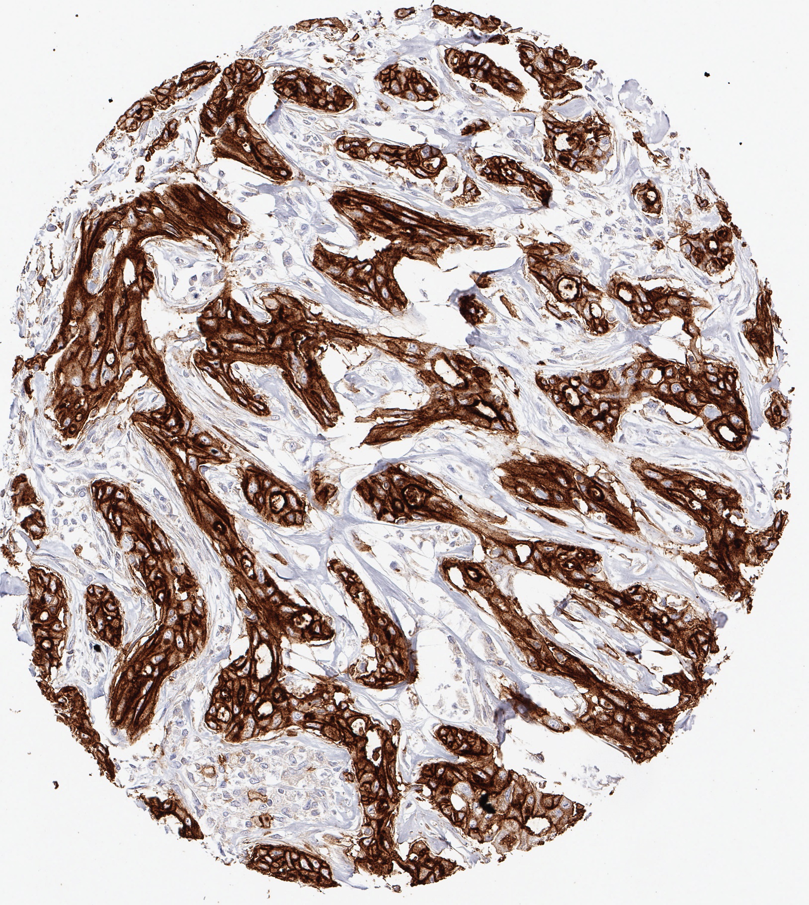
Product image of the ONCOdianova anti-CD73 antibody clone KK3: strong homogeneous CD73 immunostaining in a cholangiocellular carcinoma of the liver
Product image · Clone KK3 · DIA-KK3
Evidence layer 01

Clone-specific datasheet and protocol

Datasheet DIA-KK3 (version 25 Feb 2025/04; product content identical to version 22 Feb 2022/03) documents identity, immunogen, formulation, reconstitution, storage, IHC starting range, retrieval, detection-system-dependent dilutions, tonsil control, membranous pattern and four figures. The ONCOdianova product documentation adds a manual autoclave protocol.

Evidence layer 02

Adenosine-axis research context

CD73 IHC is being investigated in the context of adenosine-targeted therapy and patient stratification research. Clone KK3 shows membranous CD73/NT5E staining with tonsil as the documented positive control.

Evidence layer 03

Documented application and images

Brightfield IHC on human FFPE tissue, Western blot as listed on the ONCOdianova product documentation; ten gallery figures, the four datasheet figures and a manual protocol.

Brightfield and datasheet gallery: section P·03 ↓

Clone KK3 technical specifications.

ProductAnti-CD73 (Human) from Mouse — mouse monoclonal antibody; product designation of the ONCOdianova product documentation: “Anti-CD73 (HU) from Mouse (clone KK3) – 500 µl”, catalogue reference “#DIA-KK3”
CloneKK3 (monoclonal)
Catalog numberDIA-KK3 (500 µl)
Distributor catalog numberODN-DIA-KK3 (BIOZOL)
Product categoryCancer Immunology IHC marker
TargetCD73 — ecto-5′-nucleotidase (5′-nucleotidase ecto), GPI-anchored cell-membrane enzyme; gene NT5E, Gene ID 4907, protein UniProt P21589
ImmunogenPeptide of human CD73
IsotypeMouse IgG2b/κ
Host speciesMouse
ReactivityHuman
ConjugationUnconjugated
FormatLyophilized powder; antibody purified from culture supernatant; the ONCOdianova product documentation states 20 µg per vial
ReconstitutionRestore DIA-KK3 to 500 µl with sterile distilled water; gentle shaking for 10 minutes
FormulationPBS, pH 7.4, 1% BSA, 0.05% sodium azide
ApplicationsIHC on standard FFPE sections (datasheet version 25 Feb 2025/04: IHC-P) · Western blot (ONCOdianova product documentation)
IHC starting range1:100 – 1:200 (datasheet, general recommendation); in the instructions for use 1:50 for biotin/(strept)avidin-based and 1:150 for polymer-based detection; 1:150 in the manual protocol of the ONCOdianova product documentation; optimal dilution to be determined by the user for tissue, fixation and detection system
Epitope retrievalHeat-induced epitope retrieval required; autoclave at pH 7.8 recommended (Tris-EDTA-citrate, e.g. TEC buffer) (datasheet); 121 °C, 5 min in the manual protocol (ONCOdianova product documentation)
Primary antibody incubation60 min at 37 °C in the manual protocol (ONCOdianova product documentation, section P·04)
DetectionBiotin/(strept)avidin-based (e.g. ABC-HRP/AEC) or polymer-based (e.g. Dako EnVision peroxidase/DAB, 30 min at 37 °C) (datasheet); EnVision HRP rabbit/mouse, 30 min at 37 °C (ONCOdianova product documentation)
Positive controlTonsil
Staining patternMembranous
StorageLyophilized at 2–8 °C; long-term at −20 °C (stable for at least one year); reconstituted at 2–8 °C short term (several weeks); avoid repeated freeze/thaw cycles
Associated antibodyDIA-TC8 (anti-CD8, clone TC8) — as listed in the datasheet
Manufacturer / BrandONCOdianova — field “Manufacturer / Brand” of the ONCOdianova product documentation
StatusFor Research Use Only. Not for use in diagnostic procedures.

The technical specifications above are documented in datasheet DIA-KK3 (version 25 Feb 2025/04), whose product content is identical to the version of 22 Feb 2022/03, and on the ONCOdianova product documentation where indicated; gene and protein entries link to their registries. Each laboratory validates its own conditions.

IHC protocols.

Three documented application contexts: the manual ONCOdianova workflow (A), general datasheet guidance (B) and detection-specific datasheet conditions (C). Each dilution is presented with its corresponding detection system. These documented conditions provide practical starting points for laboratory establishment of KK3 IHC workflows.

A · ONCOdianova application protocolManual stain with autoclaveONCOdianova product page DIA-KK3

Source: ONCOdianova product documentation DIA-KK3, tab “IHC protocol”, headed “Staining protocols for anti-human CD73 antibody clone KK3” with the note “(will be updated continuously)”. Every value of the three protocol steps comes from that source alone.

  1. Pretreatment: TRIS-EDTA-citrate buffer (TEC buffer, Dako) — 121 °C / 5 min / pH 7.8
  2. Incubation primary antibody: 60 min / 37 °C — clone KK3 dilution 1:150
  3. EnVision HRP rabbit/mouse: 30 min / 37 °C

Positive control according to the datasheet: tonsil; expected staining pattern: membranous.

B · Datasheet — general guidanceImmunohistochemical staining of standard formalin-fixed paraffin sectionsDatasheet DIA-KK3 · V04

Source: datasheet DIA-KK3, version 25 Feb 2025/04; the instructions for use are identical in version 22 Feb 2022/03.

  1. General recommendation of the product table: dilution 1:100–1:200 (IHC-P) — general starting range of the product table
  2. Reconstitution: restore DIA-KK3 to 500 µl with sterile distilled water, gentle shaking for 10 minutes
  3. Deparaffinize and rehydrate according to standard procedures
  4. Heat-induced epitope retrieval (HIER) is required; pretreatment in an autoclave at pH 7.8 is recommended (Tris-EDTA-citrate, pH 7.8, e.g. TEC buffer)
  5. Run positive and negative controls in parallel; positive control: tonsil; expected pattern: membranous
C · Datasheet — two separate detection approachesDetection-dependent dilutionDatasheet DIA-KK3 · V04

Source: datasheet DIA-KK3, version 25 Feb 2025/04, section “Instructions for Use”.

Approach 1 — biotin/(strept)avidin-based detection

  1. Use the antibody at 1:50 — documented dilution for this detection approach
  2. Example given in the datasheet: Vectastain Elite ABC-HRP kit / AEC

Approach 2 — polymer-based detection

  1. Use the antibody at 1:150 — documented dilution for this detection approach
  2. Example given in the datasheet: Dako EnVision detection system (peroxidase/DAB), 30 min, 37 °C

Approach 1 and approach 2 are two detection approaches of the datasheet, each with its own documented dilution.

How to read the staining.

Expected pattern

Membranous CD73 staining pattern with clone KK3

The datasheet describes a membranous visualization with tonsil as positive control. The ONCOdianova figures show membranous CD73 staining of tumor cells — strong and homogeneous in a cholangiocellular carcinoma, heterogeneous in a hepatocellular carcinoma, with apical predominance in an adenocarcinoma of the lung, strong and diffuse in a squamous cell carcinoma of the lung, prominent in a malignant melanoma and strong in an angiosarcoma — as well as staining of the superficial membrane of the placental syncytiotrophoblast and CD73 staining of lymphocytes in a Warthin tumor of the parotid gland.

Documented validation scope

Product validation and documented use

Clone KK3 was developed and validated specifically for the immunohistochemical detection of CD73 in routine FFPE human tissue specimens, and is highly efficient for CD73 detection in normal and tumor FFPE tissue.

Statements of the ONCOdianova website — “Clone KK3 has been developed and validated specifically for the immunohistochemical (IHC) detection of CD73 in routine FFPE human tissue specimen.” (CD73 IHC gallery) · “Clone KK3, validated for CD73 detection in FFPE tissue.” and “Clone KK3 is highly efficient in detection of CD73-adenosine in normal and tumor FFPE tissue.” (ONCOdianova home page).

Research use

Tissue-based CD73 research

KK3 visualises CD73 protein in human FFPE sections. Researchers can assess its staining pattern and cellular distribution alongside morphology and tissue compartments. The original ONCOdianova images illustrate tissue-specific CD73 patterns in normal and tumour specimens. CD73 is described on tumor cells, cancer-associated fibroblasts and other stromal cells, endothelium and lymphocyte subsets.[5, 7, 8]

CD73/NT5E in the adenosine checkpoint.

Product information of the ONCOdianova CD73 gallery (“Clone KK3 validated for studying the CD73-adenosine checkpoint in FFPE tissues”) and of the “Reaktivität” section of the ONCOdianova product documentation.

CD73 and CD39 are cell surface enzymes that catabolize the breakdown of extracellular ATP into adenosine. As ectonucleosidases they play important roles in maintaining tissue and immune homeostasis through interfering with the extracellular purinergic pathway. Since several labs independently have demonstrated an immunosuppressive role of CD73-adenosine in cancer, the CD73-adenosine axis has emerged as one of the most promising therapeutic targets in immuno-oncology.

Adenosine triphosphate (ATP) is the major source of energy for the cell. Malignant cells can release high levels of ATP (e.g. after damage by radiotherapy or chemotherapy). Extracellular ATP provokes inflammation by driving “purinergic signals” and plays a significant role in promoting anti-tumor responses. Many tumors express the ectonucleotidases CD39 and CD73 to scavenge such proinflammatory mediators and generate immunosuppressive adenosine nucleosides.

Clone KK3 has been developed and validated specifically for the immunohistochemical (IHC) detection of CD73 in routine FFPE human tissue specimen. Given the broad expression of ectonucleotidases and adenosine receptors, immunohistochemical (IHC) application of monoclonal antibody KK3 may help to develop a better understanding of cell- and tissue-specific roles. Moreover, besides elucidation of the mechanisms-of-action of CD73 and adenosine-targeting agents it is important to differentiate patient populations. In translational research, IHC with KK3 can contribute tissue-level information to investigations of CD73- and adenosine-targeted approaches, differences between patient populations and possible associations between CD73 expression patterns and treatment response.

CD73 (ecto-5′-nucleotidase, encoded by the human NT5E gene) is a GPI-anchored cell-surface enzyme that hydrolyses extracellular nucleoside monophosphates, preferentially AMP, to the corresponding nucleoside — for AMP, adenosine. Together with CD39 (ENTPD1), which converts ATP and ADP to AMP, it forms the extracellular ATP–adenosine pathway; adenosine acts through adenosine receptors and is described as an immunosuppressive mediator in the tumor microenvironment.[1, 2, 4, 9] In experimental models, CD73 on tumor cells impaired antitumor T-cell responses,[5] and CD73 on cancer-associated fibroblasts was enhanced by an A2B-receptor-mediated feed-forward circuit.[7]

The datasheet reactivity text summarizes this context: ATP is the major energy source of the cell; malignant cells can release high levels of ATP, for example after damage by radiotherapy or chemotherapy; extracellular ATP drives purinergic signals that promote inflammation and anti-tumor responses, and many tumors express the ectonucleotidases CD39 and CD73, which remove such pro-inflammatory mediators and generate adenosine. NT5E/CD73 expression is heterogeneous across tumor types, and its prognostic associations differed between entities in the published multi-cancer analysis.[8] Anti-CD73 and adenosine-receptor approaches are subjects of immuno-oncology research.[1, 3, 6] Given the broad expression of ectonucleotidases and adenosine receptors, tissue-based immunohistochemistry can support studies of the cell- and tissue-specific distribution of CD73.

These statements describe the target CD73 and its research context. Clone-specific application data for KK3 are given in the product data, the protocols, the gallery and the documents section of this page.

  • GeneNT5E — NCBI Gene 4907, chromosome 6q14.3
  • Protein5′-nucleotidase (ecto-5′-nucleotidase, CD73), 574 amino acids — UniProt P21589
  • AliasesCD73 · NT5E · eN · eNT · NT5 · E5NT · ecto-5′-nucleotidase
  • LocalizationGPI-anchored cell-membrane enzyme — the basis of the membranous staining pattern
  • Research evaluationMembranous CD73 staining can be evaluated together with morphology, tissue compartment and complementary markers according to the study design
  • Related cloneThe datasheet lists DIA-TC8 (anti-CD8, clone TC8) as associated antibody; see also the ONCOdianova clones R12, TG1 and TG2

Bracketed numbers refer to the target-literature list below. Target biology is kept separate from clone-specific product claims.

CD73 in translational immuno-oncology research.

Target-level

CD73 translational research

CD73 participates in extracellular adenosine generation and is under therapeutic investigation. Coveler et al. (2024) evaluated CD73 expression in exploratory biomarker analyses within a randomised study of oleclumab-based combinations in metastatic pancreatic cancer. This provides a clinical-study example of CD73 biomarker research alongside investigation of adenosine-targeted treatment approaches.[10] Details: CD73 in the research overview.

Clone

What the clone can be used to study

Clone KK3 can be used to study CD73 expression on tumor and stromal cells in human FFPE tissue, documented by its datasheet with tonsil as control, a membranous staining pattern and clone-labelled figures of tonsil, placenta and tumor tissues.

Target-level

Translational research context

Published studies provide translational target-level context for CD73 research in the tumor microenvironment and in adenosine-targeted therapy studies.

Target literature and CD73 translational research.

  1. Leone RD, Emens LA. Targeting adenosine for cancer immunotherapy. Journal for ImmunoTherapy of Cancer (2018); 6(1):57. doi:10.1186/s40425-018-0360-8 · PMID 29914571Target biology · review
  2. Allard D, Chrobak P, Allard B, Messaoudi N, Stagg J. Targeting the CD73-adenosine axis in immuno-oncology. Immunology Letters (2019); 205:31–39. doi:10.1016/j.imlet.2018.05.001 · PMID 29758241Target biology · review
  3. Antonioli L, Blandizzi C, Malavasi F, Ferrari D, Haskó G. Anti-CD73 immunotherapy: A viable way to reprogram the tumor microenvironment. OncoImmunology (2016); 5(9):e1216292. doi:10.1080/2162402X.2016.1216292 · PMID 27757316Target biology · review
  4. Allard B, Beavis PA, Darcy PK, Stagg J. Immunosuppressive activities of adenosine in cancer. Current Opinion in Pharmacology (2016); 29:7–16. doi:10.1016/j.coph.2016.04.001 · PMID 27209048Target biology · review
  5. Jin D, Fan J, Wang L, Thompson LF, Liu A, Daniel BJ, Shin T, Curiel TJ, Zhang B. CD73 on tumor cells impairs antitumor T-cell responses: a novel mechanism of tumor-induced immune suppression. Cancer Research (2010); 70(6):2245–2255. doi:10.1158/0008-5472.CAN-09-3109 · PMID 20179192Target biology
  6. Chen S, Wainwright DA, Wu JD, Wan Y, Matei DE, Zhang Y, Zhang B. CD73: an emerging checkpoint for cancer immunotherapy. Immunotherapy (2019); 11(11):983–997. doi:10.2217/imt-2018-0200 · PMID 31223045Target biology · review
  7. Yu M, Guo G, Huang L, Deng L, Chang CS, Achyut BR, Canning M, Xu N, Arbab AS, Bollag RJ, Rodriguez PC, Mellor AL, Shi H, Munn DH, Cui Y. CD73 on cancer-associated fibroblasts enhanced by the A2B-mediated feedforward circuit enforces an immune checkpoint. Nature Communications (2020); 11:515. doi:10.1038/s41467-019-14060-x · PMID 31980601Target biology
  8. Jiang T, Xu X, Qiao M, Li X, Zhao C, Zhou F, Gao G, Wu F, Chen X, Su C, Ren S, Zhai C, Zhou C. Comprehensive evaluation of NT5E/CD73 expression and its prognostic significance in distinct types of cancers. BMC Cancer (2018); 18(1):267. doi:10.1186/s12885-018-4073-7 · PMID 29514610Target biology
  9. Vijayan D, Young A, Teng MWL, Smyth MJ. Targeting immunosuppressive adenosine in cancer. Nature Reviews Cancer (2017); 17(12):709–724. doi:10.1038/nrc.2017.86 · PMID 29059149
    Erratum published 2017 (Nature Reviews Cancer 17(12):765; doi:10.1038/nrc.2017.110).
    Target biology · review
  10. Coveler AL, Reilley MJ, Zalupski M, et al. A Phase Ib/II Randomized Clinical Trial of Oleclumab with or without Durvalumab plus Chemotherapy in Patients with Metastatic Pancreatic Ductal Adenocarcinoma. Clinical Cancer Research (2024); 30(20):4609–4617. doi:10.1158/1078-0432.CCR-24-0499 · PMID 39106081CD73 translational research

Entry 9 carries an erratum. Entries 1–5 are the five references of the datasheet and the ONCOdianova product documentation, cited with the bibliographic data indexed at PubMed (entry 2 with the print year 2019; entry 3 is an editorial); entries 6–9 are the review and interpretation sources of this page; entry 10 (Coveler et al. 2024) is cited as a clinical-study example of CD73 translational biomarker research. The literature provides scientific context on CD73 biology, the adenosine axis and immuno-oncology research.

Frequent scientific questions.

What is CD73 clone KK3?

KK3 is a mouse monoclonal antibody (IgG2b/κ) raised against a peptide of human CD73, the GPI-anchored ecto-5′-nucleotidase encoded by the gene NT5E. It is offered as a research reagent for immunohistochemistry on human formalin-fixed, paraffin-embedded (FFPE) tissue with a membranous staining pattern.

What is DIA-KK3?

DIA-KK3 is the ONCOdianova catalog number of the anti-CD73 antibody clone KK3: mouse monoclonal (IgG2b/κ), reactivity human, IHC on FFPE tissue, lyophilized, reconstituted to 500 µl, tonsil as positive control. The distributor catalog number at BIOZOL is ODN-DIA-KK3.

Is KK3 suitable for human FFPE IHC?

Yes. The datasheet states that clone KK3 was developed and validated specifically for the immunohistochemical detection of CD73 in routine FFPE human tissue specimens, gives a starting range of 1:100 to 1:200 and separate dilutions for biotin/(strept)avidin-based (1:50) and polymer-based detection (1:150). The ONCOdianova product documentation documents a manual autoclave protocol, and ten original ONCOdianova figures show CD73 staining in tonsil, placenta and eight tumor tissues.

What is the recommended dilution for KK3 IHC?

The datasheet gives 1:100 to 1:200 as a general recommendation for IHC on FFPE sections and, in the instructions for use, 1:50 for biotin/(strept)avidin-based detection (e.g. ABC-HRP/AEC) and 1:150 for polymer-based detection (e.g. Dako EnVision, peroxidase/DAB). The manual protocol of the ONCOdianova product documentation uses 1:150 with 60 min at 37 °C. The optimal dilution is determined by the user for tissue, fixation, platform and detection system.

Which epitope retrieval is used for KK3?

Heat-induced epitope retrieval is required. The datasheet recommends pretreatment in an autoclave at pH 7.8 (Tris-EDTA-citrate, e.g. TEC buffer); the manual protocol of the ONCOdianova product documentation specifies 121 °C for 5 min in TEC buffer, pH 7.8.

Which positive control and staining pattern are expected?

Tonsil is the positive control named in the datasheet; the expected visualization is membranous. The ONCOdianova figures show membranous CD73 staining of tumor cells in cholangiocellular carcinoma (strong, homogeneous), hepatocellular carcinoma (heterogeneous), adenocarcinoma of the lung (apical predominance), squamous cell carcinoma of the lung (strong, diffuse), malignant melanoma and angiosarcoma, staining of the superficial membrane of the placental syncytiotrophoblast, and CD73 staining of lymphocytes in a Warthin tumor of the parotid gland.

What positive control tissue is recommended for KK3?

Tonsil. It is the positive control tissue named in the KK3 datasheet, and a tonsil figure is part of the documented ONCOdianova image material.

What staining pattern does clone KK3 show?

The datasheet documents a membranous visualization. In the ONCOdianova figures the membranous CD73 staining is tissue-specific — for example strong and homogeneous in a cholangiocellular carcinoma, heterogeneous in a hepatocellular carcinoma and with apical predominance in an adenocarcinoma of the lung.

Is CD73 staining membranous?

Yes — the KK3 datasheet documents membranous visualization, consistent with CD73 being a GPI-anchored cell-membrane enzyme. The documented patterns are tissue-specific and include apical predominance and heterogeneous membranous staining.

What information does KK3 staining provide in FFPE tissue?

KK3 visualises CD73 protein in human FFPE sections. The staining pattern and cellular distribution can be assessed alongside morphology and tissue compartments; the original ONCOdianova images illustrate tissue-specific CD73 patterns in normal and tumour specimens.

Which cells and compartments can express CD73?

CD73 is expressed by many cell types. In the published literature it is described on tumor cells, on cancer-associated fibroblasts and stromal cells, on endothelium and on lymphocyte subsets, and it occurs in normal epithelial and lymphoid tissues; the ONCOdianova figures show tumor cells, syncytiotrophoblast and lymphocytes. Tumour, stromal, endothelial and immune-cell staining can be evaluated separately and, depending on the research question, together with additional markers.

What is the documented validation scope of clone KK3?

Clone KK3 was developed and validated specifically for the immunohistochemical detection of CD73 in routine FFPE human tissue specimens, with a documented protocol and figures from tonsil, placenta and eight tumor tissues; it is highly efficient for CD73 detection in normal and tumor FFPE tissue.

Which format and formulation does DIA-KK3 have?

DIA-KK3 is a lyophilized antibody purified from culture supernatant, reconstituted to 500 µl with sterile distilled water; the ONCOdianova product documentation states 20 µg per vial. The datasheet gives the formulation as PBS, pH 7.4, with 1% BSA and 0.05% sodium azide. Store lyophilized at 2–8 °C, long term at −20 °C; reconstituted at 2–8 °C for several weeks; avoid repeated freeze/thaw cycles.

What is NT5E?

NT5E is the human gene (chromosome 6q14.3, NCBI Gene 4907) encoding ecto-5′-nucleotidase, the cell-surface enzyme known by its CD name CD73. Clone KK3 detects the CD73 protein encoded by NT5E in human FFPE tissue.

Is NT5E the same as CD73?

Yes. CD73 is the protein name of ecto-5′-nucleotidase (5′-nucleotidase ecto), encoded by the human NT5E gene on chromosome 6q14.3 (NCBI Gene 4907); further aliases are eN, eNT, NT5 and E5NT. UniProt entry P21589 describes the 574-amino-acid GPI-anchored cell-membrane enzyme.

What is ecto-5′-nucleotidase?

Ecto-5′-nucleotidase is the enzyme name of CD73 (gene NT5E): a GPI-anchored cell-surface enzyme that hydrolyses extracellular AMP to adenosine. Its membrane localization is the basis of the membranous CD73 staining pattern in IHC.

Which CD73 antibody is used for NT5E detection?

CD73 and NT5E denote the same target — the NT5E gene encodes the CD73 protein. The ONCOdianova antibody for the immunohistochemical detection of this target in routine human FFPE tissue is clone KK3 (DIA-KK3). KK3 detects the CD73 protein in human FFPE tissue with a membranous staining pattern.

What is the difference between CD39 and CD73?

CD39 (ENTPD1) and CD73 (NT5E) are two distinct ectonucleotidases of the extracellular ATP–adenosine pathway: CD39 converts ATP and ADP to AMP, and CD73 hydrolyses AMP to adenosine. Clone KK3 is directed against CD73 (NT5E).

What is the role of CD73 in the adenosine pathway?

CD73 hydrolyses extracellular AMP to adenosine and thereby forms, together with CD39, the extracellular ATP–adenosine pathway. Adenosine is described in the published literature as an immunosuppressive mediator in the tumor microenvironment, and CD73 is a target of the adenosine checkpoint in immuno-oncology research.

Is CD73 used in adenosine-targeted therapy research?

CD73 IHC is being investigated in the context of adenosine-targeted therapy and patient stratification research. Clone KK3 is a research reagent for studying CD73 expression in human FFPE tissue in this field.

Which CD73 antibody clone is suitable for human FFPE tissue?

ONCOdianova clone KK3 was developed and validated specifically for the immunohistochemical detection of CD73 in routine FFPE human tissue specimens, with membranous visualization, tonsil as positive control, a documented manual protocol and original figures from tonsil, placenta and eight tumor tissues.

How does CD73 staining vary between tumor tissues?

In the documented ONCOdianova figures, the CD73 staining pattern with clone KK3 is tissue-specific: strong and homogeneous membranous staining in a cholangiocellular carcinoma, heterogeneous staining in a hepatocellular carcinoma, apical predominance in an adenocarcinoma of the lung, strong diffuse staining in a squamous cell carcinoma of the lung, prominent staining in a malignant melanoma and strong staining in an angiosarcoma. Tumor-cell, stromal and immune-cell staining can be evaluated separately according to the study design.

Where can I download the current KK3 datasheet?

The current datasheet of anti-CD73 clone KK3 (DIA-KK3), version 25 February 2025, is available as a PDF download in the Documents section of this page. Its product content is identical to the version of 22 February 2022; the update concerns the distribution address. The datasheet documents identity, immunogen, formulation, reconstitution, storage, the IHC starting range with detection-system-dependent dilutions, epitope retrieval, tonsil as positive control, the membranous staining pattern and four figures.

Ordering and catalogue information.

Datasheet

DIA-KK3

Product datasheet, version 25 Feb 2025/04 (product content identical to the version of 22 Feb 2022/03; updated distribution address). Identity, immunogen, formulation, reconstitution, storage, instructions for use, four figures and the reference list.

Download PDF ↓

PDF · version 2025/04 · 225 KB

IHC gallery

CD73 IHC gallery

The ONCOdianova “CD73 IHC-Gallery” is presented in section P·03 of this page with ten figures and their legends, plus the datasheet figures.

Go to gallery ↓

11 figures · brightfield, datasheet

Protocol

IHC protocol

The manual autoclave protocol of the ONCOdianova product documentation and the instructions for use of the datasheet are documented in section P·04.

Go to protocols ↓

ONCOdianova protocol · datasheet guidance · section P·04

Safety

MSDS

Material safety data sheet (ONCOdianova, all lyophilized antibodies).

Download PDF ↓

PDF · V02 · 216 KB

Ordering. ONCOdianova products are purchased through our distribution partner BIOZOL Diagnostica Vertrieb GmbH, Oehleckerring 11–13, 22419 Hamburg, Germany — order requests by e-mail: order@biozol.de (CC info@oncodianova.com). See also order information.

The technical data on this page are taken from datasheet DIA-KK3 (version 25 Feb 2025/04; product content identical to the version of 22 Feb 2022/03) and from the ONCOdianova product documentation where indicated. Related: R12 (anti-CD112R/PVRIG) · TG1 (anti-TIGIT) · TG2 (anti-TIGIT) · TC8 (anti-CD8) · FX3 (FOXP3).

For Research Use Only. Not for use in diagnostic procedures.