TC8 — anti-CD8 for tumour-infiltrating lymphocytes in brightfield and multiplex IHC.

Clone TC8 · Cat. no. DIA-TC8 · T-cell co-receptor CD8, membranous staining pattern

Scientific USP

5 clone-specific peer-reviewed publications

2,652Evaluable tumors
84Tumor entities
3,988Tumors · CD8/Ki67 multiplex · digital quantification
  • Brightfield + multiplex IHC
  • Membranous CD8 · human FFPE

Clone TC8 displays highest signal-to-noise contrast and has been validated for detection of CD8+ TILs in multiplex assays.

Clone TC8 validated for studying tumor infiltrating CD8 positive T cells in FFPE tissues.

ONCOdianova home page and CD8 gallery · Datasheet 25 Feb 2025/08 · IHC gallery · Protocols · Publications — cohort sizes as reported in the respective clone-specific studies.

TC8 (DIA-TC8) is a mouse monoclonal anti-CD8 antibody (clone TC8, ONCOdianova GmbH) developed and validated for the immunohistochemical detection of CD8 in routine human FFPE tissue, for studying CD8-positive lymphocytes in the tumour microenvironment. Documented with tonsil as positive control, six staining protocols for automated platforms and manual procedures, and 36 original ONCOdianova figures from 17 tumour types. Clone TC8 was used in five peer-reviewed studies covering several thousand human tumour samples in brightfield and Opal multiplex IHC with digital quantification. Documented applications: IHC on FFPE tissue (datasheet), fluorescence multiplex IHC (ONCOdianova home page and product page) and western blot (ONCOdianova product documentation).

  • Host / IsotypeMouse · IgG2a/κ
  • ReactivityHuman
  • ApplicationsIHC on FFPE tissue · multiplex fluorescence IHC · Western blot
  • IHC starting range1:100 – 1:200
  • Positive controlTonsil · membranous
  • Format500 µl (DIA-TC8) · 100 µl (DIA-TC8-M), lyophilized
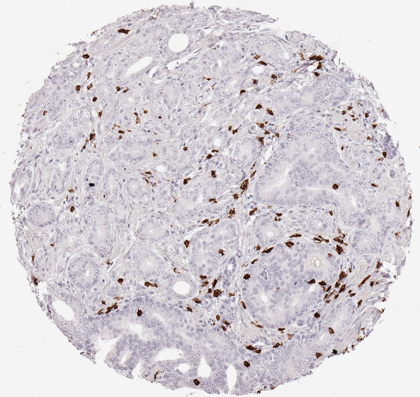
Product image of the ONCOdianova anti-CD8 antibody clone TC8: CD8 immunohistochemistry showing a dense infiltrate of CD8-positive lymphocytes in adenocarcinoma of the prostate
Product image · Clone TC8 · DIA-TC8
Evidence layer 01

Clone-specific datasheet and protocols

Datasheet DIA-TC8 (version 25 Feb 2025/08; product content identical to version 22 Feb 2022/09) documents identity, immunogen, formulation, reconstitution, storage, IHC starting range, tonsil control, membranous pattern, three automated platforms and four figures. The ONCOdianova product documentation adds six staining protocols.

Evidence layer 03

Documented applications and images

Brightfield IHC and fluorescence multiplex IHC on human FFPE tissue, Western blot as listed on the ONCOdianova product documentation; 36 gallery figures, the product image of the ONCOdianova product documentation, the four datasheet figures and one CD8 × CD112R/PVRIG multiplex of tonsil in which the anti-CD8 clone is named.

Brightfield, datasheet and multiplex gallery: section P·03 ↓

Controlled product data.

ProductAnti-CD8 (Human) from Mouse — mouse monoclonal anti-T cell marker (cytotoxic T cells), clone TC8 (datasheet title)
CloneTC8 (monoclonal)
Catalog numbersDIA-TC8 (500 µl) · DIA-TC8-M (100 µl) — both listed in the datasheet; the ONCOdianova product documentation lists DIA-TC8, 500 µl
Distributor catalog numberODN-DIA-TC8 (BIOZOL)
Product categoryCancer Immunology IHC marker
TargetCD8 — T-cell surface glycoprotein CD8, co-receptor of the T-cell receptor on cytotoxic T cells (datasheet: CD8). Registry entries: α chain CD8A, Gene ID 925, UniProt P01732; β chain CD8B, Gene ID 926, UniProt P10966
ImmunogenRecombinant peptide of human CD8
IsotypeMouse IgG2a/κ
Host speciesMouse
ReactivityHuman
ConjugationUnconjugated
FormatLyophilized powder; antibody purified from culture supernatant; the ONCOdianova product documentation states 100 µg per vial
ReconstitutionRestore DIA-TC8 to 500 µl (DIA-TC8-M to 100 µl) with sterile distilled water; gentle shaking for 10 minutes
FormulationPBS, pH 7.4, 1% BSA, 0.05% sodium azide
ApplicationsIHC on standard FFPE sections (datasheet: IHC-P) · fluorescence multiplex IHC (ONCOdianova product documentation and home page; two clone-specific publications) · Western blot (ONCOdianova product documentation)
IHC starting range1:100 – 1:200 (datasheet, general recommendation); 1:200 in the platform protocols and 1:100 in the manual microwave protocol of the ONCOdianova product documentation; 1:200 and 1:450 in the published studies (study-specific); optimal dilution to be determined by the user for tissue, fixation, platform and detection system
Epitope retrievalHeat-induced epitope retrieval required (datasheet); pH 9 in the platform protocols (Dako Autostainer Link 48: 15 min / 95 °C; Leica Bond RX: 15 min / 100 °C; Ventana Discovery Ultra: 24 min / 100 °C), pH 7.8 by autoclave (121 °C, 5 min) or microwave in the manual protocols (ONCOdianova product documentation)
Primary antibody incubationDako Autostainer Link 48: 20 min; Leica Bond RX: 15 min; Ventana Discovery Ultra: 60 min / 38 °C; manual protocols: 60 min / 37 °C (ONCOdianova product documentation, section P·04)
Positive controlTonsil
Staining patternMembranous (CD8-positive lymphocytes)
Automated platformsVentana Discovery Ultra, Leica Bond RX, Dako Autostainer Link 48 (datasheet); platform programs on the ONCOdianova product documentation; Dako Autostainer Link 48 also in two published studies
StorageLyophilized at 2–8 °C; long-term at −20 °C (stable for at least one year); reconstituted at 2–8 °C short term (several weeks); avoid repeated freeze/thaw cycles
Associated antibodiesDIA-TG1 (anti-TIGIT, clone TG1) · DIA-R12 (anti-CD112R/PVRIG, clone R12) — as listed in the datasheet
Alternative namesCD8, Cluster of Differentiation 8, T-cell surface glycoprotein CD8 alpha chain, T-lymphocyte differentiation antigen T8/Leu-2, Leu2, Leu2 T lymphocyte antigen, T cell antigen Leu2, T8 T cell antigen, MAL, OKT8 T cell antigen, T cell co receptor, CD8a, CD8A — list of the distributor BIOZOL (ODN-DIA-TC8)
Manufacturer / BrandONCOdianova GmbH
StatusFor Research Use Only. Not for use in diagnostic procedures.

The technical specifications above are documented in datasheet DIA-TC8 (version 25 Feb 2025/08), whose product content is identical to the version of 22 Feb 2022/09, and on the ONCOdianova product documentation where indicated; gene and protein entries link to their registries. Each laboratory validates its own conditions.

IHC protocols and published applications.

ONCOdianova protocol

Documented ONCOdianova protocols — six workflows

Six staining workflows are documented for clone TC8: three automated platforms, two manual procedures (cards below) and a manual two-colour immunofluorescence (block B). Each dilution is presented with its corresponding workflow.

Dako Autostainer Link 48
Pretreatment
15 min · 95 °C · pH 9
Primary antibody
1:200 · 20 min · room temperature
Linker
no
Detection
HRP (polymer) · 20 min · room temperature
Leica Bond RX
Pretreatment
15 min · 100 °C · pH 9
Primary antibody
1:200 · 15 min
Post Primary
8 min
Detection
HRP (polymer) · 8 min
Ventana Discovery Ultra
Pretreatment
24 min · 100 °C · pH 9
Primary antibody
1:200 · 60 min · 38 °C
Secondary antibody
12 min · 36 °C
Detection
HRP (polymer) · 12 min
Manual — autoclave
Pretreatment
121 °C · 5 min · pH 7.8
Primary antibody
1:200 · 60 min · 37 °C
Detection
EnVision HRP rabbit/mouse · 30 min · 37 °C
Manual — microwave
Pretreatment
90 s at 1000 W to boiling, then 15 min · 270 W · pH 7.8
Primary antibody
1:100 · 60 min · 37 °C
Detection
EnVision HRP rabbit/mouse · 30 min · 37 °C

Source: ONCOdianova product page DIA-TC8, tab “IHC protocols” — “Staining protocols for anti-human CD8 antibody clone TC8”. The values apply per workflow; datasheet DIA-TC8 gives 1:100–1:200 as the starting range and is kept separate in block A below.

External published application

Manual brightfield IHC — Fraune et al.

Antibody
Clone TC8
Dilution
1:450
Workflow
manual
Antigen retrieval
Autoclave · 121 °C · 5 min · Tris-EDTA-citrate · pH 7.8
Primary incubation
60 min at 37 °C
Detection
EnVision Kit (Dako)

Fraune C. et al., Annals of Surgical Oncology 2020;27(10):3997–4006 · doi:10.1245/s10434-020-08209-y

External published application

Automated brightfield IHC — Blessin et al. 2020

Antibody
Clone TC8
Dilution
1:200
Antigen retrieval
DAKO PT-LINK · 98 °C · 15 min · Target Retrieval Solution S2367 · pH 9
Platform
DAKO Link 48
Peroxidase block
5 min
Primary incubation
20 min at room temperature
Detection
Flex HRP 20 min · DAB 10 min · haematoxylin 5 min

Blessin N.C. et al., Cellular Oncology 2020;43(3):421–430 · doi:10.1007/s13402-020-00496-7

External published application

Multiplex fluorescence IHC — Blessin et al. 2021

CD8
Oncodianova · clone TC8 · Cat# DIA-TC8
Antigen retrieval
pH 9.0
Dilution
1:200
Staining position
2
Opal dye
Opal 690

Blessin N.C. et al., Aging 2021;13(11):14590–14603 · doi:10.18632/aging.203113 — table 5, CD8 row.

The protocols and published workflows shown here are documented application examples from their respective sources; the documented conditions provide practical starting points for laboratory establishment of DIA-TC8 workflows.

Three documented application contexts: the current datasheet (A), the six protocols of the ONCOdianova product documentation for automated platforms and manual procedures (B) and the conditions reported in the clone-specific studies (C). Each dilution is presented with its corresponding workflow and detection system. These documented conditions provide practical starting points for laboratory establishment of TC8 IHC workflows.

A · DatasheetImmunohistochemical staining of standard formalin-fixed paraffin sectionsDatasheet DIA-TC8 · version 25 Feb 2025/08 (identical wording in version 22 Feb 2022/09)

Preparation and epitope retrieval

  1. Reconstitution: restore DIA-TC8 to 500 µl with sterile distilled water (DIA-TC8-M to 100 µl), gentle shaking for 10 minutes
  2. Deparaffinize and rehydrate according to standard procedures
  3. Heat-induced epitope retrieval (HIER) is required

Primary antibody and detection

  1. Use the antibody at 1:100–1:200 (IHC-P, general recommendation)
  2. Detection alternatives: indirect immunoenzyme labeling with a secondary antibody conjugate, biotin/(strept)avidin-based, soluble enzyme immune complex or polymer-based
  3. Suited for automated platforms (datasheet: Ventana Discovery Ultra, Leica Bond RX, Dako Autostainer Link 48); the platform programs are given in block B
  4. Run positive and negative controls in parallel; positive control: tonsil; expected pattern: membranous

For IHC protocols the datasheet refers to www.oncodianova.com (block B).

B · ONCOdianova product documentationSix staining protocols: Dako, Leica, Ventana, manual (autoclave, microwave), two-colour immunofluorescenceONCOdianova product page DIA-TC8, section “IHC protocols” (as of 30 Aug 2026)

Dako Autostainer Link 48

  1. Pretreatment buffer: 15 min / 95 °C / pH 9
  2. Incubation primary antibody: 20 min / room temperature, dilution 1:200; linker: no
  3. HRP (polymer): 20 min / room temperature

Leica Bond RX

  1. Pretreatment buffer: 15 min / 100 °C / pH 9
  2. Incubation primary antibody: 15 min, dilution 1:200
  3. Post Primary: 8 min; HRP (polymer): 8 min

Ventana Discovery Ultra

  1. Pretreatment buffer: 24 min / 100 °C / pH 9
  2. Incubation primary antibody: 60 min / 38 °C, dilution 1:200
  3. Secondary antibody: 12 min / 36 °C; HRP (polymer): 12 min

Manual stain with autoclave

  1. Pretreatment buffer: 121 °C / 5 min / pH 7.8
  2. Incubation primary antibody: 60 min / 37 °C, dilution 1:200
  3. EnVision HRP rabbit/mouse: 30 min / 37 °C

Manual stain with microwave

  1. Pretreatment buffer: 90 s at 1000 W to boiling, then 15 min / 270 W / pH 7.8
  2. Incubation primary antibody: 60 min / 37 °C, dilution 1:100
  3. EnVision HRP rabbit/mouse: 30 min / 37 °C

Two-colour immunofluorescence, manual stain

  1. Position 1: antibody at pH 9; position 2: CD8
  2. Pretreatment buffer: 90 s at 1000 W to boiling, then 15 min / 270 W / pH 9
  3. Incubation primary antibody: 30 min / room temperature, dilution 1:200; HRP (polymer): 10 min / room temperature

The product page additionally states the reconstitution to 500 µl.

C · Published study conditionsBrightfield IHC (autostainer and manual) and Opal multiplex — published application contextsThree studies with TC8-specific conditions (methods sections)

C1 · Automated brightfield IHC, Dako Autostainer Link 48 (Blessin et al. 2020)

  1. FFPE tissue microarrays; epitope retrieval PT Link, pH 9, 15 min at 98 °C
  2. Peroxidase block 5 min; “Oncodianova, mouse monoclonal antibody, Clone TC8, dilution 1:200”, 20 min at room temperature
  3. EnVision Flex HRP 20 min, DAB 10 min, haematoxylin 5 min; digitization Leica Aperio VERSA 8 (40×), analysis with HALO or ImageScope, CD8-positive cells/mm²

C2 · Manual brightfield IHC (Fraune et al. 2020, pancreatic carcinoma)

  1. Epitope retrieval: autoclave 5 min at 121 °C, Tris-EDTA-citrate buffer pH 7.8
  2. “Oncodianova, mouse monoclonal antibody, Clone TC8, 1:450”, 60 min at 37 °C; Dako EnVision; digital cell counting

C3 · Opal multiplex immunofluorescence (Blessin et al. 2021, Aging)

  1. Position 2: CD8 “Oncodianova, Clone: TC8, Cat#: DIA-TC8”, retrieval pH 9.0, 1:200, Opal 690 (table 5)

The 1:200 and 1:450 dilutions belong to the respective described procedures (platform, retrieval, incubation time and detection); the datasheet starting range is 1:100–1:200. The publications name “Oncodianova” as supplier.

How to read the staining.

Expected pattern

Membranous, CD8-positive lymphocytes

The datasheet describes a membranous visualization with tonsil as positive control. The ONCOdianova figures show CD8-positive lymphocytes as scattered, moderate or dense infiltrates in the tumour stroma, at the stroma–epithelium interface or between tumour cells, in carcinomas of 14 organs, in a neuroendocrine tumour of the pancreas, in colon adenoma and in thymoma, without staining of the tumour cells in the depicted fields. In the published normal-tissue panel of Blessin et al. 2020, splenic blood vessels were the only clearly CD8-positive non-lymphatic cell type.

Documented validation scope

Product validation and documented use

Clone TC8 was developed specifically for the routine immunohistochemical detection of CD8 in FFPE tissue and validated for the identification of CD8-positive tumour-infiltrating T cells (TILs) in order to allow the detection of CD8 in the tumour microenvironment, including validation for the detection of CD8-positive TILs in multiplex assays and an optimization of the signal-to-noise contrast. Five peer-reviewed studies independently used clone TC8 in brightfield IHC on 2,652 tumours of 84 entities with 608 normal tissues, on 1,163 renal tumours and on 551 pancreatic carcinomas, and DIA-TC8 in Opal multiplex IHC with Ki67 on 3,988 tumours of six cancer types and on normal, inflammatory and colorectal tissue, each with digital quantification.

Research use

Research interpretation of CD8 staining

TC8 visualises the CD8 co-receptor on the surface of CD8-positive lymphocytes in human FFPE sections. CD8-positive T cells are the classical cytotoxic effector cells,[1] and tumour-infiltrating CD8-positive T cells comprise phenotypically distinct populations, including bystander cells.[2] Researchers can evaluate the density and spatial distribution of CD8-positive lymphocytes together with morphology and tissue compartment and, according to the research question, with further markers — the published multiplex studies combined TC8 with Ki67 to assess the proliferation of CD8-positive cells.

CD8 in research.

Reactivity text of datasheet DIA-TC8 (version 25 Feb 2025/08; identical wording in version 22 Feb 2022/09).

Clone TC8 has been developed specifically for routine immunohistochemical (IHC) detection of CD8 in formalin-fixed paraffin-embedded tissue specimen. TC8 has been validated for the identification of CD8 positive tumor infiltrating T cells (TILs) in order to allow the detection of CD8 in the tumor microenvironment under pathological conditions.

The T-cell receptor (TCR) recognizes specific antigenic peptides on the surface of cancer and other target cells presented by HLA-I/β2m complexes. Binding to TCR induces a signalling transduction cascade, leading to execution of cytotoxic T lymphocyte (CTL) functions. Thus, CD8+ T cells are directly involved in antitumor cytotoxic responses, while inhibitory T-cell receptors such as PD-1, CTLA-4 and TIGIT are activated by the immunosuppressive tumor microenvironment with the aim to inactivate tumor-infiltrating lymphocytes (TILs). The most effective current cancer immunotherapies include immune checkpoint inhibition ICI and block T-cell inhibitory receptors. Moreover, effective blockade immunotherapy appears to be associated with the presence of CD8+ T cells.

Product information of the ONCOdianova CD8 gallery and of the “Reactivity” section of the ONCOdianova product documentation.

CD8 antibody Clone TC8 has been developed specifically for the immunohistochemical (IHC) detection of CD8 in routine FFPE human tissue specimen. Clone TC8 has been optimized for optimal signal to noise contrast and validated for the identification of CD8+ tumor infiltrating T cells (TILs) with the aim to allow an unequaled specific detection of CD8 in the tumor microenvironment.

CD8+ T cells play a central role for the killing of cancer cells. They have the ability to infiltrate different human tumors and are engaged in the development of a specific tumor microenvironment. Cancer cells have developed mechanisms to successfully evade the antitumor immune response by generating inhibitory signals through upregulation of the expression of immunosuppressive components. Effective blockade of this interaction is considered as a major factor in the development of cancer immunotherapies. Moreover, preexisting CD8+ T cells seem to predict the efficacy of such immune checkpoint therapies.

The T-cell receptor (TCR) recognizes specific antigenic peptides on the surface of cancer and other target cells presented by HLA-I/β2m complexes. Binding to TCR induces a signaling transduction cascade, leading to execution of cytotoxic T lymphocyte (CTL) functions. While CD8+ T cells are directly involved in antitumor cytotoxic responses, the involvement of CD4+ T cells is more indirect, e.g. by their help in priming of CD8+ T cells.

In contrast, inhibitory T-cell receptors such as PD-1, CTLA-4 and TIGIT are activated by the immunosuppressive tumor microenvironment with the aim to inactivate tumor-infiltrating lymphocytes (TILs). The most effective current cancer immunotherapies include immune checkpoint inhibition ICI and block T-cell inhibitory receptors. Moreover, effective blockade immunotherapy appears to be associated with the presence of CD8+ T cells.

ONCOdianova product documentation, “Reactivity”: Clone TC8 has been developed specifically for the immunohistochemical (IHC) detection of CD8 in routine FFPE human tissue specimen. TC8 has been validated for the identification of CD8 positive tumor infiltrating T cells (TILs) with the aim to allow an unequaled specific detection of CD8 in the tumor microenvironment. IHC application of monoclonal antibody TC8 may provide valuable information for clinical research and potential therapeutic interventions targeting the tumor immunology checkpoint.

CD8 is a cell-surface glycoprotein and co-receptor of the T-cell receptor. On conventional cytotoxic T cells it is expressed as a CD8αβ heterodimer encoded by the genes CD8A and CD8B; CD8αα homodimers occur on further lymphocyte populations. The co-receptor binds MHC class I and supports the recognition of peptide antigens presented by MHC class I molecules, the basis of the cytotoxic T-cell response.[1, 7] Tumour-specific cytotoxic T-cell clones have been derived from lung cancer patients,[3, 4, 5] and the activation of CD8-positive T cells is controlled by regulatory T cells[6] and by the immunosuppressive tumour microenvironment.[9]

In tissue studies, CD8 immunohistochemistry is used to determine the density and spatial distribution of CD8-positive lymphocytes in tumours: effector memory T cells in colorectal cancer were associated with early metastasis and survival in the published cohort,[8] and pre-existing CD8-positive T cells at the tumour margin were associated with responses to PD-1 blockade in the published melanoma cohort.[11] Neoantigen-directed T-cell responses and their broadening under checkpoint therapy are subjects of current research.[10, 12] The datasheet reactivity text summarizes this context: inhibitory receptors such as PD-1, CTLA-4 and TIGIT are activated in the immunosuppressive tumour microenvironment, and effective checkpoint blockade appears to be associated with the presence of CD8-positive T cells.

These statements describe the target CD8 and its research context. Clone-specific application data for TC8 are given in the product data, the protocols, the gallery, the publications and the documents section of this page.

  • GenesCD8A — NCBI Gene 925 · CD8B — NCBI Gene 926
  • ProteinsT-cell surface glycoprotein CD8 α chain — UniProt P01732 · β chain — UniProt P10966
  • AliasesCD8 · Leu-2 · T8 · CD8α (p32) · CD8β (p37)
  • LocalizationCell-surface co-receptor — the basis of the membranous staining pattern
  • Research evaluationCD8-positive lymphocytes can be evaluated by density and spatial distribution together with morphology, tissue compartment and complementary markers according to the study design; tumour-infiltrating CD8-positive T cells comprise phenotypically distinct populations — Simoni et al. 2018 [2]
  • Related clonesCD8 × CD112R/PVRIG multiplex documented with R12; see also the ONCOdianova anti-TIGIT clones TG1 and TG2

Bracketed numbers refer to the target-literature list below. Target biology is kept separate from clone-specific product claims.

CD8 in translational immuno-oncology research.

Target-level

CD8 translational research

CD8 is a central marker for cytotoxic T-cell infiltration. Pre-existing CD8-positive T cells at the tumour margin were associated with responses to PD-1 blockade in the published melanoma cohort of Tumeh et al. (2014),[11] and CD8 IHC has been used in paired pretreatment and on-treatment biopsies of immunotherapy trials, where changes in CD8-positive T-cell density were associated with clinical outcome in published trial analyses. Details: CD8 in the research overview.

Clone

What the clone can be used to study

Clone TC8 can be used to study CD8-positive tumor-infiltrating lymphocytes and their density in human FFPE tissue — in automated brightfield IHC with digital quantification and in Opal multiplex immunofluorescence — and is named in five peer-reviewed studies on several thousand human FFPE tumors.

Target-level

Translational research context

Published studies provide translational target-level context for CD8 research in the tumour microenvironment and in immunotherapy studies.

Clone-specific evidence and target literature.

  1. Blessin NC, Spriestersbach P, Li W, Mandelkow T, Dum D, Simon R, Hube-Magg C, Lutz F, Viehweger F, Lennartz M, Fraune C, Nickelsen V, Fehrle W, Göbel C, Weidemann S, Clauditz T, Lebok P, Möller K, Steurer S, Izbicki JR, Sauter G, Minner S, Jacobsen F, Luebke AM, Büscheck F, Höflmayer D, Wilczak W, Burandt E, Hinsch A. Prevalence of CD8+ cytotoxic lymphocytes in human neoplasms. Cellular Oncology (2020); 43(3):421–430. doi:10.1007/s13402-020-00496-7 · PMID 32141029 · PMC7214387
    Automated brightfield IHC (Dako Autostainer Link 48, PT Link pH 9, 1:200, 20 min at room temperature, EnVision Flex/DAB) on tissue microarrays: 2,652 evaluable tumours of 84 tumour entities and 608 normal tissues of 76 tissue types; digital quantification of CD8-positive cells per mm² (Aperio VERSA 8, HALO).
    Peer-reviewed article · clone TC8
  2. Eichenauer T, Simmendinger L, Fraune C, Mandelkow T, Blessin NC, Kluth M, Hube-Magg C, Möller K, Clauditz T, Weidemann S, Dahlem R, Fisch M, Riechardt S, Simon R, Sauter G, Büscheck F, Rink M. High level of EZH2 expression is linked to high density of CD8-positive T-lymphocytes and an aggressive phenotype in renal cell carcinoma. World Journal of Urology (2021); 39(2):481–490. doi:10.1007/s00345-020-03200-4 · PMID 32303902 · PMC7910252
    Automated brightfield IHC (Dako Autostainer Link 48, PT Link pH 9, 1:200, 20 min at room temperature, EnVision Flex/DAB) on a tissue microarray of 1,809 renal tumours, 1,163 interpretable for CD8; digital quantification (Aperio VERSA 8, ImageScope).
    Peer-reviewed article · clone TC8
  3. Blessin NC, Abu-Hashem R, Mandelkow T, Li W, Simon R, Hube-Magg C, Möller-Koop C, Witt M, Schmidt A, Büscheck F, Fraune C, Luebke AM, Möller K, Jacobsen F, Lutz F, Lennartz M, Steurer S, Sauter G, Höflmayer D, Tsourlakis MC, Hinsch A, Burandt E, Wilczak W, Minner S, Clauditz TS. Prevalence of proliferating CD8+ cells in normal lymphatic tissues, inflammation and cancer. Aging (2021); 13(11):14590–14603. doi:10.18632/aging.203113 · PMID 34083496 · PMC8221353
    Opal multiplex immunofluorescence CD8 × Ki67 on FFPE sections (CD8: DIA-TC8, retrieval pH 9.0, 1:200, position 2, Opal 690 — table 5): normal lymphatic tissues, inflammatory tissues, tumours and 765 evaluable colorectal carcinomas; quantification with ImageScope/HALO.
    Peer-reviewed article · DIA-TC8
  4. Blessin NC, Li W, Mandelkow T, Jansen HL, Yang C, Raedler JB, Simon R, Büscheck F, Dum D, Luebke AM, Hinsch A, Möller K, Menz A, Bernreuther C, Lebok P, Clauditz T, Sauter G, Marx A, Uhlig R, Wilczak W, Minner S, Krech T, Fraune C, Höflmayer D, Burandt E, Steurer S. Prognostic role of proliferating CD8+ cytotoxic Tcells in human cancers. Cellular Oncology (2021); 44(4):793–803. doi:10.1007/s13402-021-00601-4 · PMID 33864611 · PMC8338812
    Opal multiplex immunofluorescence CD8 × Ki67 on FFPE sections (CD8: Cat# DIA-TC8, clone TC8, 1:200): 3,988 evaluable colorectal, breast, renal cell, ovarian, pancreatic and gastric carcinomas; quantification with HALO.
    Peer-reviewed article · DIA-TC8
  5. Fraune C, Burandt E, Simon R, Hube-Magg C, Makrypidi-Fraune G, Kluth M, Büscheck F, Höflmayer D, Blessin NC, Mandelkow T, Li W, Perez D, Izbicki JR, Wilczak W, Sauter G, Schrader J, Neipp M, Mofid H, Daniels T, Isbert C, Clauditz TS, Steurer S. MMR Deficiency is Homogeneous in Pancreatic Carcinoma and Associated with High Density of Cd8-Positive Lymphocytes. Annals of Surgical Oncology (2020); 27(10):3997–4006. doi:10.1245/s10434-020-08209-y · PMID 32108923 · PMC7471097
    Manual brightfield IHC (autoclave 121 °C, 5 min, Tris-EDTA-citrate pH 7.8; 1:450, 60 min at 37 °C; Dako EnVision) on tissue microarrays of 597 pancreatic carcinomas, 551 evaluable for CD8; digital cell counting. Correction published 2022 (doi:10.1245/s10434-022-11798-5): an acknowledgement was added; methods and results unchanged.
    Peer-reviewed article · clone TC8

Bibliographic data as indexed at PubMed (entry 2, Eichenauer et al., cited with the print year 2021; the ONCOdianova product documentation cites its online publication of 2020). The five studies originate from the pathology research environment of the University Medical Center Hamburg-Eppendorf and document the published use of clone TC8 in automated brightfield IHC, manual brightfield IHC and Opal multiplex immunofluorescence under the stated conditions.

  1. Blessin NC, Mandelkow T, Bady E, Hube-Magg C, Sauter G, Simon R, Fraune C, Lennartz M, Weidemann SA, Möller K, Höflmayer D. Abstract 3870: Patterns of CD112R expression in normal lymphatic tissues, inflammation and cancer. Cancer Research (Proceedings AACR Annual Meeting 2020) (2020); 80(16 Suppl):3870. doi:10.1158/1538-7445.AM2020-3870Congress abstract
  2. Simon R, Blessin NC, Mandelkow T, Bady E, Hube-Magg C, Sauter G, Lennartz M, Fraune C, Weidemann SA, Möller K, Büscheck F. Abstract 4970: Prognostic role of CD112R, PD-1 and Ki67 expression in CD8+cytotoxic T cells in colorectal cancer. Cancer Research (Proceedings AACR Annual Meeting 2020) (2020); 80(16 Suppl):4970. doi:10.1158/1538-7445.AM2020-4970Congress abstract

The datasheet and the ONCOdianova product documentation list these two AACR 2020 abstracts among the “specific references for clone TC8”. Both abstracts describe CD112R multiplex immunohistochemistry with CD8 and Ki67 as co-markers from the same Hamburg research environment; they are listed here as the references cited by the datasheet, and the peer-reviewed clone-specific studies above document the published use of clone TC8.

  1. Zhang N, Bevan MJ. CD8(+) T cells: foot soldiers of the immune system. Immunity (2011); 35(2):161–168. doi:10.1016/j.immuni.2011.07.010 · PMID 21867926Target biology · review
  2. Simoni Y et al. Bystander CD8+ T cells are abundant and phenotypically distinct in human tumour infiltrates. Nature (2018); 557(7706):575–579. doi:10.1038/s41586-018-0130-2 · PMID 29769722Target biology
  3. Weynants P, Thonnard J, Marchand M, Delos M, Boon T, Coulie PG. Derivation of tumor-specific cytolytic T-cell clones from two lung cancer patients with long survival. American Journal of Respiratory and Critical Care Medicine (1999); 159(1):55–62. doi:10.1164/ajrccm.159.1.9805073 · PMID 9872818Target biology
  4. Echchakir H, Vergnon I, Dorothée G, Grunenwald D, Chouaib S, Mami-Chouaib F. Evidence for in situ expansion of diverse antitumor-specific cytotoxic T lymphocyte clones in a human large cell carcinoma of the lung. International Immunology (2000); 12(4):537–546. doi:10.1093/intimm/12.4.537 · PMID 10744655Target biology
  5. Karanikas V et al. High frequency of cytolytic T lymphocytes directed against a tumor-specific mutated antigen detectable with HLA tetramers in the blood of a lung carcinoma patient with long survival. Cancer Research (2001); 61(9):3718–3724. PMID 11325844Target biology
  6. Piccirillo CA, Shevach EM. Cutting edge: control of CD8+ T cell activation by CD4+CD25+ immunoregulatory cells. Journal of Immunology (2001); 167(3):1137–1140. doi:10.4049/jimmunol.167.3.1137 · PMID 11466326Target biology
  7. Bossi G, Trambas C, Booth S, Clark R, Stinchcombe J, Griffiths GM. The secretory synapse: the secrets of a serial killer. Immunological Reviews (2002); 189:152–160. doi:10.1034/j.1600-065x.2002.18913.x · PMID 12445272Target biology · review
  8. Pagès F et al. Effector memory T cells, early metastasis, and survival in colorectal cancer. New England Journal of Medicine (2005); 353(25):2654–2666. doi:10.1056/NEJMoa051424 · PMID 16371631Target biology
  9. Gajewski TF, Meng Y, Harlin H. Immune suppression in the tumor microenvironment. Journal of Immunotherapy (2006); 29(3):233–240. doi:10.1097/01.cji.0000199193.29048.56 · PMID 16699366Target biology · review
  10. Kvistborg P et al. Anti-CTLA-4 therapy broadens the melanoma-reactive CD8+ T cell response. Science Translational Medicine (2014); 6(254):254ra128. doi:10.1126/scitranslmed.3008918 · PMID 25232180Target biology
  11. Tumeh PC et al. PD-1 blockade induces responses by inhibiting adaptive immune resistance. Nature (2014); 515(7528):568–571. doi:10.1038/nature13954 · PMID 25428505Target biology
  12. Schumacher TN, Schreiber RD. Neoantigens in cancer immunotherapy. Science (2015); 348(6230):69–74. doi:10.1126/science.aaa4971 · PMID 25838375Target biology · review

Entries 1 and 2 are the review and interpretation sources of this page; entries 3–12 are the ten “general references” of the ONCOdianova product documentation, cited with the bibliographic data indexed at PubMed. The literature provides scientific context on CD8 biology, tumour-infiltrating lymphocytes and immuno-oncology research.

Frequent scientific questions.

What does anti-CD8 clone TC8 detect?

TC8 is a mouse monoclonal antibody (IgG2a/κ) raised against a recombinant peptide of human CD8, the T-cell co-receptor of the T-cell receptor. It is offered as a research reagent for immunohistochemistry on human formalin-fixed, paraffin-embedded (FFPE) tissue with a membranous staining pattern of CD8-positive lymphocytes.

Is TC8 documented for FFPE immunohistochemistry?

Yes. The datasheet states that clone TC8 was developed specifically for the routine immunohistochemical detection of CD8 in FFPE tissue specimens and gives a starting range of 1:100 to 1:200. The ONCOdianova product documentation documents six protocols (Dako Autostainer Link 48, Leica Bond RX, Ventana Discovery Ultra, manual autoclave, manual microwave, two-colour immunofluorescence), and 36 original ONCOdianova figures show CD8-positive lymphocytes in 17 tumour types.

Are there peer-reviewed publications using clone TC8?

Yes. Five peer-reviewed studies used clone TC8 (catalogue number DIA-TC8): Blessin et al. 2020 (Cellular Oncology; 2,652 tumours of 84 entities and 608 normal tissues), Eichenauer et al. 2021 (World Journal of Urology; renal cell carcinoma), Blessin et al. 2021 (Aging; CD8 × Ki67 multiplex in normal, inflammatory and tumour tissue), Blessin et al. 2021 (Cellular Oncology; CD8 × Ki67 multiplex in 3,988 tumours of six cancer types) and Fraune et al. 2020 (Annals of Surgical Oncology; pancreatic carcinoma).

Is TC8 validated for fluorescence multiplex IHC?

Yes. Clone TC8 was validated for the detection of CD8-positive TILs in multiplex assays; the ONCOdianova product documentation gives a manual two-colour immunofluorescence protocol with CD8 in position 2, and datasheet DIA-R12 shows a CD8 × CD112R/PVRIG multiplex of normal tonsil with DIA-TC8. Independently published evidence includes two peer-reviewed Opal multiplex studies (Blessin et al. 2021, Aging and Cellular Oncology) that used DIA-TC8 at 1:200 together with Ki67 (Aging, table 5: position 2, Opal 690). Each panel is established by the user for tissue, antibody order, retrieval, dilution and detection.

Which dilution should be used?

The datasheet gives 1:100 to 1:200 as a general recommendation for IHC on FFPE sections. The platform protocols of the ONCOdianova product documentation use 1:200 (Dako, Leica, Ventana, manual autoclave, two-colour immunofluorescence) and 1:100 for the manual microwave protocol. Published studies used 1:200 (Dako Autostainer Link 48 and Opal multiplex) and 1:450 (manual autoclave protocol, 60 min at 37 °C). Dilution, retrieval and incubation belong together; the optimal dilution is determined by the user for tissue, fixation, platform and detection system.

Which epitope retrieval is used for TC8?

Heat-induced epitope retrieval is required (datasheet). The platform protocols use pH 9 (15 min at 95 °C for Dako Autostainer Link 48, 15 min at 100 °C for Leica Bond RX, 24 min at 100 °C for Ventana Discovery Ultra); the manual protocols use pH 7.8 with autoclave (121 °C, 5 min) or microwave. Published studies used PT Link pH 9 (15 min at 98 °C) for the automated brightfield protocol, autoclave pH 7.8 for the manual protocol and pH 9.0 for the Opal multiplex protocol.

Which positive control and staining pattern are expected?

Tonsil is the positive control named in the datasheet; the expected pattern is membranous staining of CD8-positive lymphocytes. The ONCOdianova figures show CD8-positive lymphocytes within tumour stroma and between tumour cells in carcinomas of the prostate, lung, oesophagus, stomach, ovary, vulva, cervix, vagina, kidney, urinary bladder, liver, anus, pancreas, thyroid and oral cavity, in colon adenoma and in thymoma.

Can TC8 be used on automated staining platforms?

Yes. The datasheet states suitability for the Ventana Discovery Ultra, Leica Bond RX and Dako Autostainer Link 48, and the ONCOdianova product documentation gives a program for each platform (section P·04). Two published studies used the Dako Autostainer Link 48 with PT Link retrieval; the published multiplex stainings were performed manually with Opal reagents.

What information does CD8 staining with clone TC8 provide?

CD8 immunostaining with clone TC8 visualises the CD8 co-receptor on the surface of CD8-positive lymphocytes in human FFPE sections. Their density and spatial distribution — in the tumour stroma, at the stroma–epithelium interface or between tumour cells — can be evaluated together with morphology and tissue compartment; the published multiplex studies combined TC8 with Ki67 to assess the proliferation of CD8-positive cells, and further markers can be added according to the research question.

What is the documented validation scope of clone TC8?

Clone TC8 was developed specifically for the routine IHC detection of CD8 in FFPE tissue and validated for the identification of CD8-positive tumour-infiltrating T cells in order to allow the detection of CD8 in the tumour microenvironment, including validation for CD8-positive TILs in multiplex assays. Independently published clone-specific evidence includes five peer-reviewed studies with several thousand human FFPE tumour samples in brightfield and Opal multiplex IHC with digital quantification.

Which format and formulation does DIA-TC8 have?

DIA-TC8 is a lyophilized antibody purified from culture supernatant, reconstituted to 500 µl with sterile distilled water; the datasheet additionally lists DIA-TC8-M, reconstituted to 100 µl. The datasheet gives the formulation as PBS, pH 7.4, with 1% BSA and 0.05% sodium azide; the ONCOdianova product documentation states 100 µg per vial. Store lyophilized at 2–8 °C, long term at −20 °C; reconstituted at 2–8 °C for several weeks; avoid repeated freeze/thaw cycles.

Data package.

Datasheet

DIA-TC8

Product datasheet, version 25 Feb 2025/08 (product content identical to the version of 22 Feb 2022/09; updated distribution address). Identity, immunogen, formulation, reconstitution, storage, instructions for use, four figures and the reference list.

Download PDF ↓

PDF · version 2025/08 · 216 KB

IHC gallery

CD8 IHC gallery

The ONCOdianova “CD8 IHC-Gallery” is presented in section P·03 of this page with 36 figures and their legends, plus the product image of the ONCOdianova product documentation, the datasheet figures and one multiplex figure.

Go to gallery ↓

39 figures · brightfield, product image, datasheet, multiplex

Protocols

IHC protocols

The six staining protocols of the ONCOdianova product documentation (Dako Autostainer Link 48, Leica Bond RX, Ventana Discovery Ultra, manual autoclave, manual microwave, two-colour immunofluorescence) are documented in section P·04.

Go to protocols ↓

6 protocols · section P·04

Safety

MSDS

Material safety data sheet (ONCOdianova, all lyophilized antibodies).

Download PDF ↓

PDF · V02 · 216 KB

Ordering. ONCOdianova products are purchased through our distribution partner BIOZOL Diagnostica Vertrieb GmbH, Oehleckerring 11–13, 22419 Hamburg, Germany — order requests by e-mail: order@biozol.de (CC info@oncodianova.com). See also order information.

The technical data on this page are taken from datasheet DIA-TC8 (version 25 Feb 2025/08; product content identical to the version of 22 Feb 2022/09) and from the ONCOdianova product documentation where indicated. Related: R12 (anti-CD112R/PVRIG) · TG1 (anti-TIGIT) · TG2 (anti-TIGIT) · FX3 (FOXP3) · KK3 (CD73).

For Research Use Only. Not for use in diagnostic procedures.