
Develop
Our idea is to integrate immunohistochemistry as early as possible in the hybridoma screening procedure and thereby expand the classical ELISA screen by histopathological expertise at an early stage of FFPE antibody development.

01 / Antibody intelligence, made actionable
Monoclonal research antibodies for researchers, pathologists and biomarker teams who need evidence they can inspect.
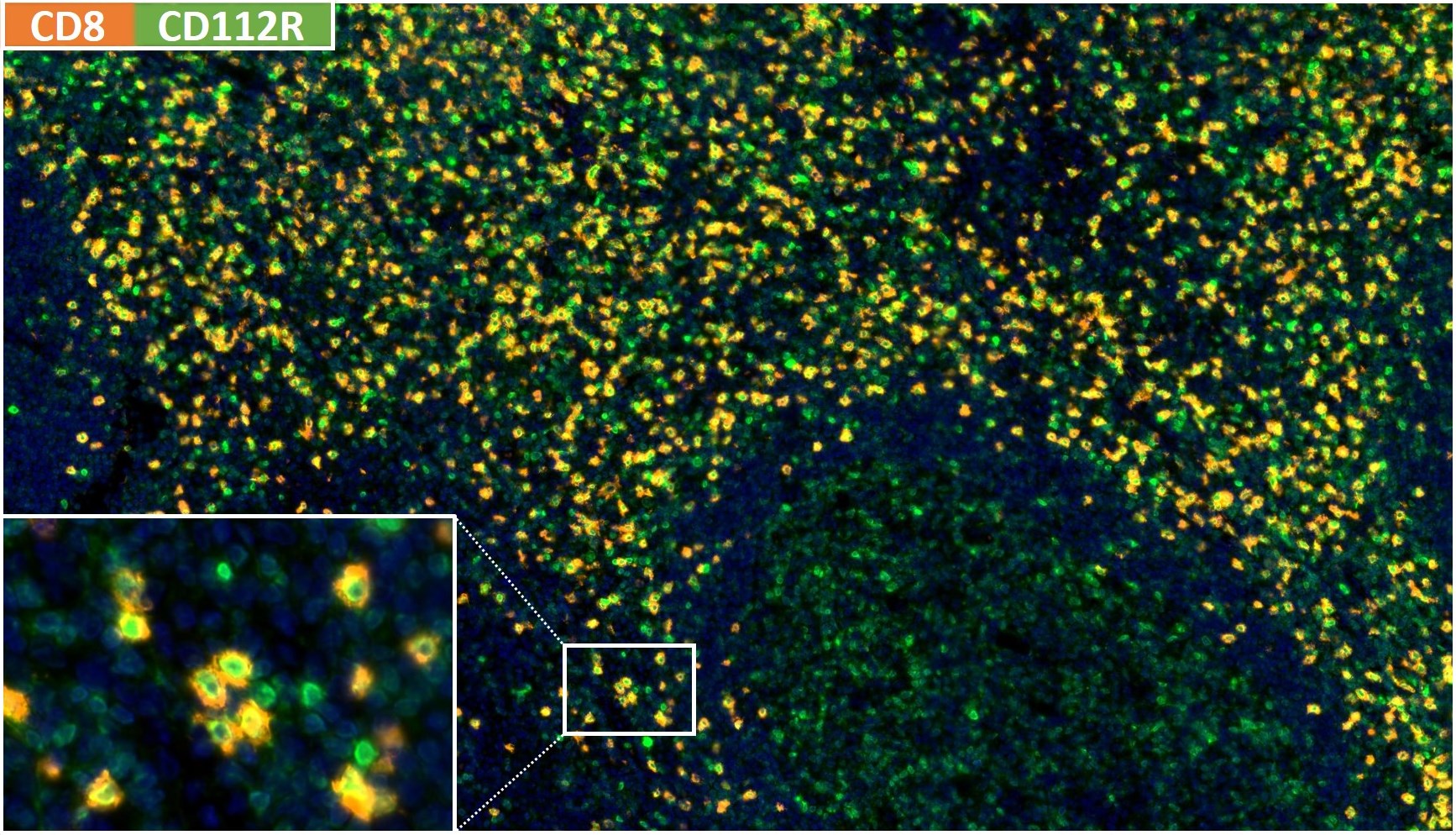
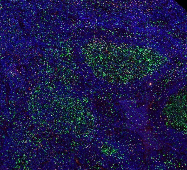
CD8 × CD112R multiplexHuman tonsil · Clones TC8 + R12 · Fluorescence IHC
For Research Use Only. Not for use in diagnostic procedures.
02 Scientific finder
Six monoclonal clones for tissue-based immuno-oncology research — searchable by target, clone and application.
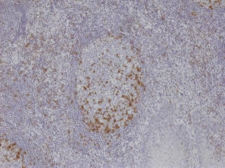 TIGITClone TG1 · DIA-TG1-MClone TG1 allows the identification of TIGIT positive T-cells in standard FFPE human tumors. TG1 is a valuable tool for clinical research of the tumor microenvironment.Clone-specifically published mouse monoclonal anti-TIGIT antibody for human FFPE, brightfield and fluorescence multiplex IHC. RRID AB_3750391.Product page
 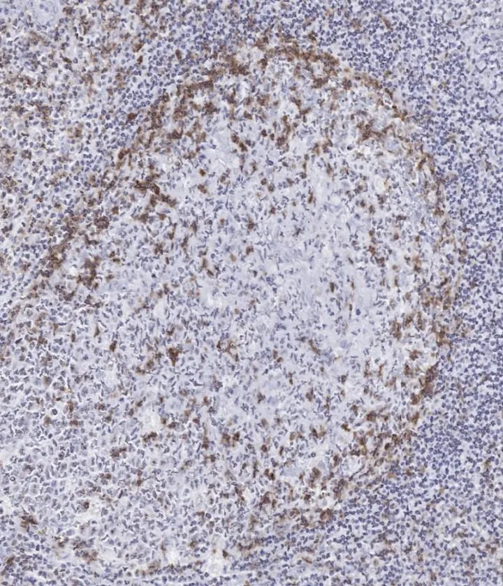 TIGITClone TG2 · DIA-TG2-MSeparate anti-TIGIT clone for IHC on human FFPE tissue.Product page
 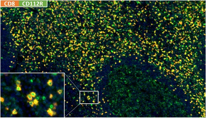 CD112R / PVRIGClone R12 · DIA-R12Clone R12 is ideally suited for multiplexed IHC studies of PVRIG/CD112R. R12 has been validated on large quantities of normal and tumor tissues.Anti-CD112R/PVRIG antibody used in a peer-reviewed multiplex immunofluorescence study (Yang et al., Modern Pathology 2023).Product page
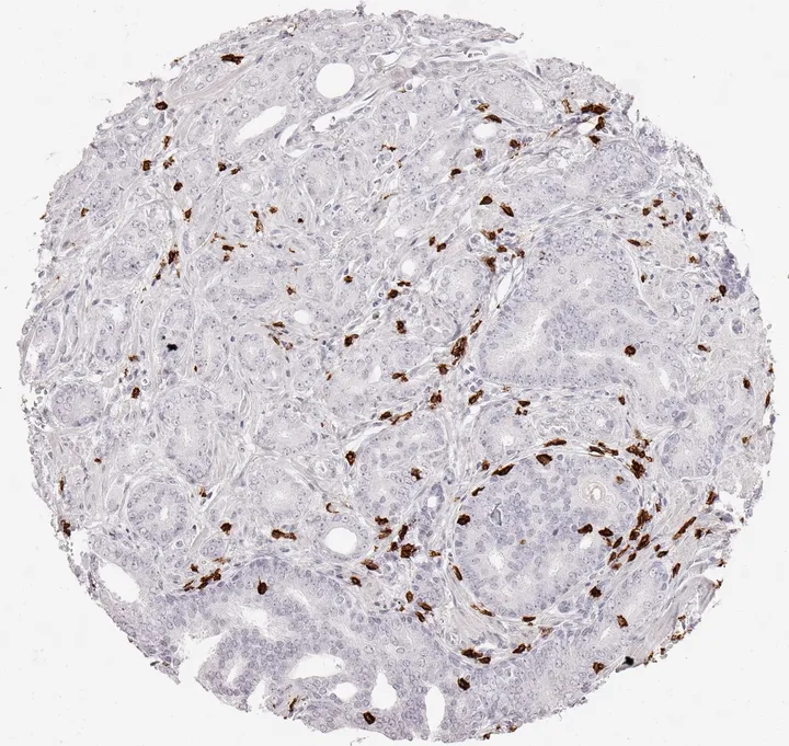
  FOXP3Clone FX3 · DIA-FX3Clone FX3 detects FOXP3 positive TILs in FFPE human tumors.Nuclear FOXP3 detection in human FFPE tissue; used in a peer-reviewed automated multiplex IF study (Picard et al., Acta Neuropathologica 2023).Product page
  CD8Clone TC8 · DIA-TC8Clone TC8 detects tumor infiltrating lymphocytes (TILs).Anti-CD8 antibody with five peer-reviewed clone-specific publications on large human tumor cohorts.Product page
 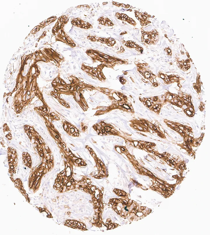 CD73Clone KK3 · DIA-KK3Clone KK3, validated for CD73 detection in FFPE tissue.Mouse monoclonal anti-CD73 (NT5E) antibody for IHC on routine human FFPE tissue; membranous staining pattern, tonsil as positive control; ten original ONCOdianova tissue figures.Product page
 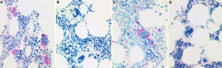 Mutant CALR (calreticulin)Clone CAL2® · dedicated siteCAL2® — mutation-specific antibody for mutant CALR. Dedicated scientific information, methodology, evidence and ordering information are available at cal2antibody.com. For Research Use Only. Not for use in diagnostic procedures.Explore CAL2®
 No marker matches this search.
03 Antibody highlights
The following antibody highlights from ONCOdianova have already developed relevance for clinical histopathology:
TIGIT
Clone TG1 · DIA-TG1-M
TIGIT is one of the most important checkpoint markers for the development of new immunotherapies against cancer. With antibody clone TG1, TIGIT could for the first time be detected immunohistochemically in tumor tissue.
Product pagePVRIG / CD112R
Clone R12 · DIA-R12
PVRIG / CD112R plays an important role in the development of effective combination therapies with PD1 and TIGIT. The IHC detection of PVRIG in tumor tissues so far is only possible with clone R12.
Product pageExplore the new anti-TIGIT clone TG2 now with higher staining intensity.
Explore TG2Our strong feedback from laboratories applying the new ONCOdianova antibodies against CD112R, TIGIT and CALRmut shows that they are perceived as innovative tools for immunohistochemistry, because they allow new approaches for tissue based studies of important biomarkers.
04 How we work
ONCOdianova is an antibody manufacturing network. The purpose is to manufacture excellent antibodies with optimized signal-to-noise ratio. Such optimized antibodies are especially needed in multicolor fluorescence based immunohistochemistry if many different antibodies are simultaneously analyzed. The mission of ONCOdianova is to develop a portfolio of “best in class antibodies” for proteins that are potentially relevant for triggering treatment decisions or for diagnostic pathology.
For Research Use Only. Not for use in diagnostic procedures.
Our idea is to integrate immunohistochemistry as early as possible in the hybridoma screening procedure and thereby expand the classical ELISA screen by histopathological expertise at an early stage of FFPE antibody development.

We use our strong network for histopathological validation of new targets in human tissues. This expands our expertise to develop new test systems for novel targets. For cancer immunology targets we put a focus on tumor infiltrating lymphocytes.

Multiplex Assay technology develops fast and depends on the availability of reliable antibody reagents. Our vision is to provide the best cancer immunology marker antibodies for combined use to improve the understanding of patient subpopulations.

Our objective is to potentially develop multiple clones for the same target by approaching one target at diverse immunogenic sites. We are able to reference mapped epitopes and open for beta testing opportunities with pharmaceutical partners.
05 Validation framework
Every product page documents peer-reviewed clone-specific evidence, congress abstracts and technical specifications, with references, catalog numbers and RRIDs where assigned.
Request a data packageContext-led clone evaluation with immunohistochemistry integrated early in development.
Pattern assessment across human normal and tumor tissues with defined positive controls.
Clone-specific use documented in peer-reviewed studies where available — each cited with DOI on the product page.
Products are organised around the questions research teams actually ask: immune phenotype, checkpoint axes, spatial arrangement and tumour context.
CD8 × CD112R multiplex · Human tonsil · Clones TC8 + R12
06 Ordering & next steps
Orders are handled through BIOZOL Diagnostica Vertrieb GmbH. Every route maps a specialist question to a product, a document or a conversation.