What is clone R12, and what does DIA-R12 identify?
R12 is a mouse monoclonal antibody (IgG1/κ) raised against a recombinant peptide of human CD112R, also called PVRIG (gene PVRIG). DIA-R12 is the ONCOdianova catalogue number of this antibody; the distributor catalogue number at BIOZOL is ODN-DIA-R12. The antibody is supplied as 100 µl lyophilisate (PBS, 2 % BSA, sodium azide) and is documented for immunohistochemistry on human formalin-fixed, paraffin-embedded (FFPE) tissue in brightfield IHC and fluorescence multiplex IHC, with tonsil as positive control.
What is the difference between CD112, CD112R, PVRIG and NECTIN2?
PVRIG and CD112R are two names for the same molecule: an inhibitory immune-checkpoint receptor of the poliovirus receptor/nectin family (gene PVRIG, formerly C7orf15) that is expressed in T-cell and NK-cell biology. CD112 and NECTIN2 (formerly PVRL2) are likewise two names for one molecule: the ligand of this receptor, expressed on antigen-presenting cells and tumor cells. Receptor and ligand are distinct proteins encoded by different genes. CD112R/PVRIG binds CD112 and competes with the costimulatory receptor CD226 (DNAM-1) for this ligand (Zhu et al. 2016). Clone R12 is directed against the receptor CD112R/PVRIG.
Is R12 suitable for immunohistochemistry on human FFPE tissue (IHC-P)?
Yes. Immunohistochemistry on formalin-fixed, paraffin-embedded sections (IHC-P) is the core documented application of clone R12. The datasheet specifies heat-induced epitope retrieval, a starting dilution of 1:100 to 1:200 for polymer-based detection, tonsil as positive control and a membranous staining pattern of lymphocyte subsets; the ONCOdianova product documentation documents a manual protocol with 1:150 and EnVision detection. Seven brightfield figures in the gallery on this page show CD112R staining with clone R12 in human FFPE tissue.
Which publications document the use of clone R12?
One peer-reviewed journal article used clone R12 (catalogue number DIA-R12): Yang et al., Modern Pathology 2023 (PMID 36788088), a fluorescence multiplex IHC study of 523 colorectal cancers. Two doctoral theses of the University of Hamburg used DIA-R12: Yang (2021) on CD112R and PD-1 in colorectal cancer and Jansen (2020) on CD112R expression in carcinomas compared with normal tissues; both are academic theses cited with their repository records. Two AACR 2020 congress abstracts of the same Hamburg group, listed as specific references on the ONCOdianova product documentation, describe CD112R multiplex IHC and are cited as congress abstracts. All entries with links are listed under Publications.
Is R12 documented for multiplex immunofluorescence (mIF)?
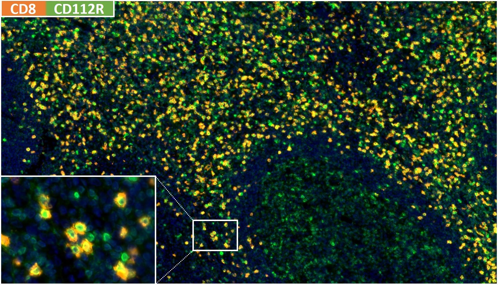
Yes. Fluorescence multiplex IHC, also termed multiplex immunofluorescence (mIF), is the published clone-specific application of DIA-R12: Yang et al. 2023 used the antibody within an Opal panel (antigen retrieval pH 7.8, dilution 1:500, Opal 570) on FFPE colorectal cancer tissue, and the theses of Yang (2021) and Jansen (2020) describe Opal-based multiplex workflows with DIA-R12. The datasheet reactivity text describes clone R12 as developed for brightfield IHC and moreover for multicolor immunofluorescence, and the ONCOdianova gallery documents CD112R × CD8 and CD112R × FOXP3 multiplex staining of human tonsil. The published conditions belong to their study panels and provide documented starting points for panel establishment.
Can R12 be combined with CD8 or FOXP3 in a multiplex panel?
Yes, both combinations are documented. A published fluorescence multiplex panel combined CD112R (clone R12) with CD8, PD-1 and Ki67 on FFPE colorectal cancer tissue (Yang et al. 2023), and the Jansen thesis (2020) evaluated CD112R on CD8+, CD4+, FOXP3+, CD56+, CD11c+ and CD68+ cells. ONCOdianova figures document CD112R × CD8 multiplex staining with clones R12 and TC8 and CD112R × FOXP3 multicolor immunofluorescence with clone R12 of normal human tonsil. Antibody order, retrieval, dilution and detection are established per panel; the published conditions provide a documented starting point.
What dilution conditions are documented for R12?
Three documented values, each within its own workflow: the datasheet states 1:100 to 1:200 for polymer-based peroxidase/DAB detection on FFPE sections (brightfield IHC-P); the manual protocol of the ONCOdianova product documentation used 1:150 with EnVision HRP detection; the published multiplex study used 1:500 within an Opal panel with Opal 570 (Yang et al. 2023). Each laboratory establishes its working dilution for its tissue, fixation, platform and detection system on this documented basis.
What antigen-retrieval conditions are documented for R12?
Heat-induced epitope retrieval is required. The datasheet recommends an autoclave at 121 °C for 5 minutes in Tris-EDTA-citrate buffer, pH 7.8 (TEC buffer); the manual protocol of the ONCOdianova product documentation uses the same pretreatment (121 °C, 5 min, pH 7.8). In the published multiplex study, the R12 step used antigen retrieval at pH 7.8 (Yang et al. 2023); the Jansen thesis (2020) describes autoclave retrieval at pH 9 within its Opal workflow. Details are given in the protocol section.
Which positive-control tissue and staining pattern are documented for R12?
Tonsil is the positive-control tissue listed for clone R12, with a membranous staining pattern of lymphocyte subsets (datasheet). Figure A of the datasheet, shown as figure 03 in the gallery, presents normal human tonsil with numerous CD112R/PVRIG-positive lymphocytes in brightfield IHC; the multiplex figures of the gallery include CD112R × CD8 and CD112R × FOXP3 staining of normal human tonsil. In the Jansen thesis (2020), DIA-R12 showed a consistently membranous pattern; the published studies report CD112R signal on CD8+, CD4+, FOXP3+ and CD56+ lymphocytes.
What evidence supports the characterization and tissue staining of clone R12?
ONCOdianova product information (datasheet reactivity text, ONCOdianova product documentation and gallery) states that clone R12 was validated for sensitivity and specificity on large quantities of normal and tumor tissues, displays no background in non-lymphoid cells and in epithelial cells, and combines high specificity with an inherently high signal-to-noise ratio, which makes it well suited for multiplex IHC studies of CD112R in human tissue. Beyond the manufacturer, the Jansen thesis (2020) reports that the specificity of DIA-R12 was examined by Western blot, ELISA and pre-absorption with CD112R protein before multiplex use, and Yang et al. 2023 used DIA-R12 in a peer-reviewed multiplex panel. Brightfield IHC, fluorescence multiplex IHC and Western blot are the documented applications.
Is Western blot a documented application of R12?
Yes. ONCOdianova's original R12 product information lists Western blot among the antibody's applications, alongside immunohistochemistry on human FFPE sections. In addition, the doctoral thesis of Jansen (2020, University of Hamburg) reports that the specificity of DIA-R12 was examined by Western blot, ELISA and pre-absorption with CD112R protein.
Is R12 suitable for automated IHC platforms?
The datasheet states that the antibody is suited for immunohistochemical staining on automated platforms, and the ONCOdianova product documentation describes its manual protocol as applicable to automated staining procedures. The documented conditions — heat-induced epitope retrieval at pH 7.8 and 1:100 to 1:200 for polymer-based detection — are the starting point for establishing R12 on a laboratory's own platform.